This site uses tracking information. Visit our privacy policy. Click to agree to this policy and not see this again.

Ophthalmology and Visual Sciences

Epithelial downgrowth

39-year-old Caucasian man referred for blurred vision and corneal edema in right eye following multiple ocular surgeries.

Epithelial downgrowth

39-year-old Caucasian man referred for blurred vision and corneal edema in right eye following multiple ocular surgeries.
Lauren Colwell, BA; Ryan J. Diel, MD; Matthew Benage, MD; Lai Jiang, MD; Mark A. Greiner MD; Jennifer J. Ling, MD
Posted February 26, 2020

INITIAL PRESENTATION

Chief Complaint

Blurry vision, right eye

History of Present Illness

A 39-year-old male with a history of uncharacterized syndromic pigmentary retinopathy, optic atrophy, childhood cataracts, and multiple ocular surgeries was referred for blurred vision and persistent corneal edema in the right eye for the past three to four months.

In his early 20s, he underwent cataract extraction with intraocular lens (CEIOL) implantation in both eyes followed by Nd:YAG laser capsulotomies. He had good vision (~20/60) for many years until the IOL in the right eye dislocated. He underwent pars-plana vitrectomy (PPV), IOL explantation, and scleral-fixation of a secondary IOL (SS-IOL) approximately 4 months prior to presentation. Since that time, he had noted blurred vision and was found to have corneal edema on follow up exams. He denied any pain, fluctuation in symptoms throughout the day, or any improvement in vision with sodium chloride 5% ophthalmic solution (Muro128).

Past Ocular History

  • Syndromic pigmentary retinopathy, both eyes
  • Optic nerve atrophy, both eyes
  • Childhood cataracts, both eyes
  • Pseudophakia, both eyes
  • Dislocated intraocular lens posteriorly, right eye s/p PPV, IOL explantation, and scleral-fixation of a secondary IOL

Past Medical History

  • Sensorineural hearing loss, bilateral (related to syndromic pigmentary retinopathy)

Pertinent Past Surgical History

  • CEIOL, both eyes (~2000)
  • Nd:YAG capsulotomy, both eyes
  • PPV, IOL explantation, SS-IOL, right eye (May 2019)

Medications

Sodium chloride 5% ophthalmic solution (Muro128) three times daily, both eyes

  • Timolol 0.5% twice daily, both eyes
  • Systane artificial tears twice daily, both eyes

Allergies (Reaction)

  • Dorzolamide (blurry vision)
  • Brimonidine (drowsiness)

Family History

Non-contributory

Social History

  • Computer server administrator
  • Never smoker
  • Occasional alcohol use
  • No illicit drug use 

Review of Systems

  • Negative except for what is detailed in the history of present illness

OCULAR EXAMINATION

Visual Acuity with correction (Snellen)

  • Right eye (OD): 20/800
  • Left eye (OS): 20/50 -2

Manifest Refraction

  • OD: Sphere +3.50, add +3.00
  • OS: Sphere +3.00, add +3.00

Ocular Motility/Alignment

  • Both eyes (OU): Full, ortho

Intraocular Pressure (IOP) by Tonopen

  • OD: 9 mmHg
  • OS: 10 mmHg

Pachymetry

  • OD: 778 microns
  • OS: 700 microns

Pupils

  • OD: 5mm (dark) → 5mm (light), irregular shape, no relative afferent pupillary defect (RAPD)
  • OS: 4mm (dark) → 3mm (light), irregular shape, no RAPD

Confrontation visual fields

  • OD: Total superior temporal deficiency; Partial outer inferior temporal, superior nasal deficiencies
  • OS: Partial outer superior temporal superior nasal, inferior nasal deficiencies 

External

  •  Normal OU

Slit lamp exam

  • Lids/lashes: Normal OU
  • Conjunctiva/sclera: Clear and quiet OU
  • Cornea:
    • OD: Superotemporal grey sheet along the endothelium with scalloped edges, extending from the temporal full-thickness clear corneal incision site; overlying mild stromal edema not involving the visual axis (Figures 1 and 2)
    • OS: Early band keratopathy nasally, pigment on endothelium, moderate guttae
  • Anterior chamber:
    • OD: Deep with 4+ pigmented cell 
    • OS: Deep and quiet
  • Iris: 360 degrees of prominent peripheral transillumination defects OU
  • Lens:
    • OD: 3-piece scleral-fixated IOL, centered
    • OS: Posterior chamber IOL, pseudophacodonesis
  • Anterior vitreous:
    • OD: Optically empty
    • OS: Normal

Color slit lamp photograph (broad beam) of the right eye demonstrating a grey sheet with scalloped edges extending along the endothelium with overlying mild corneal edema.

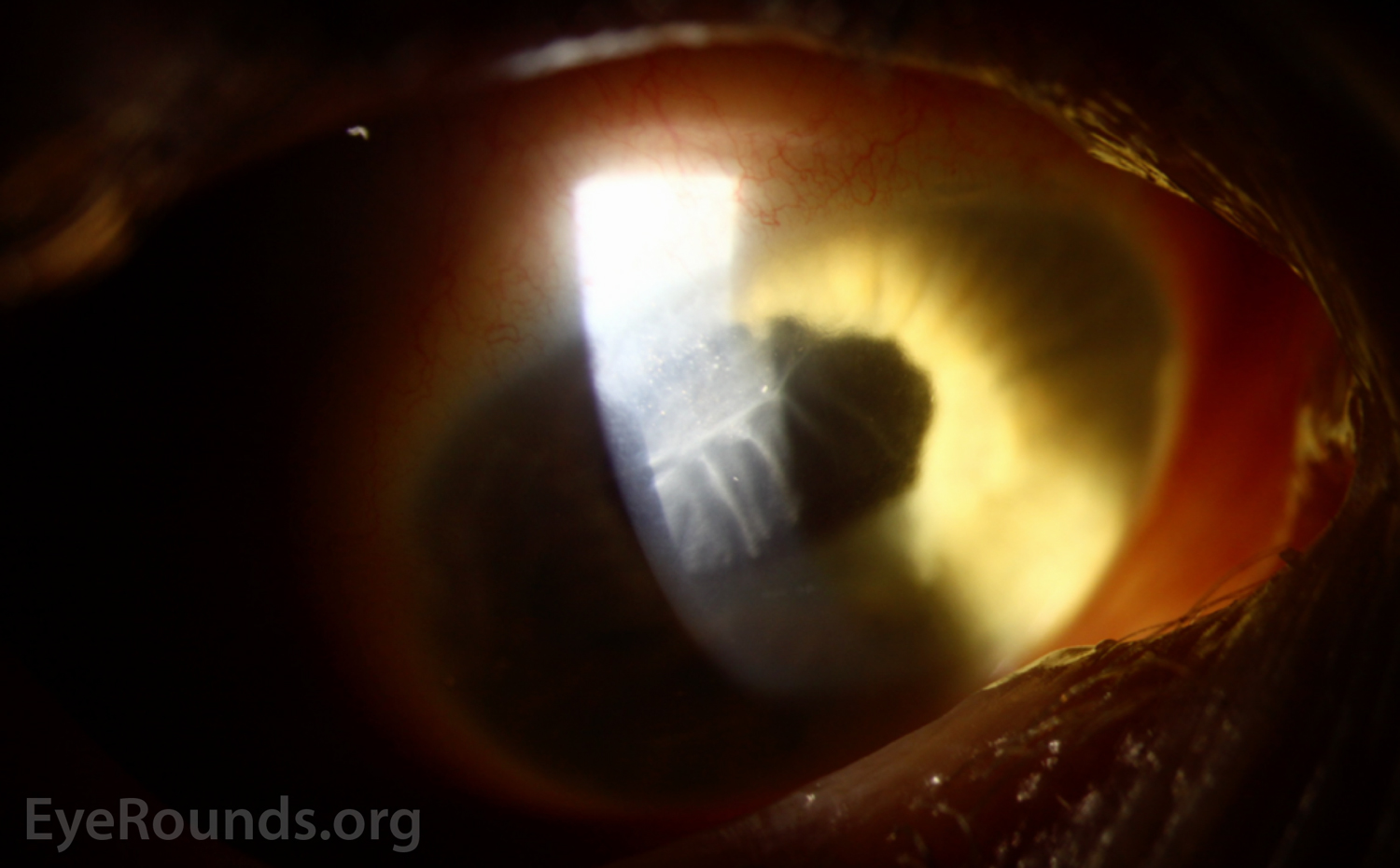
Figure 1: Color slit lamp photograph (broad beam) of the right eye demonstrating a grey sheet with scalloped edges extending along the endothelium with overlying mild corneal edema.

Color slit lamp photograph (narrow beam) of the right eye demonstrating hyperreflective cells extending along the clear corneal incision site.

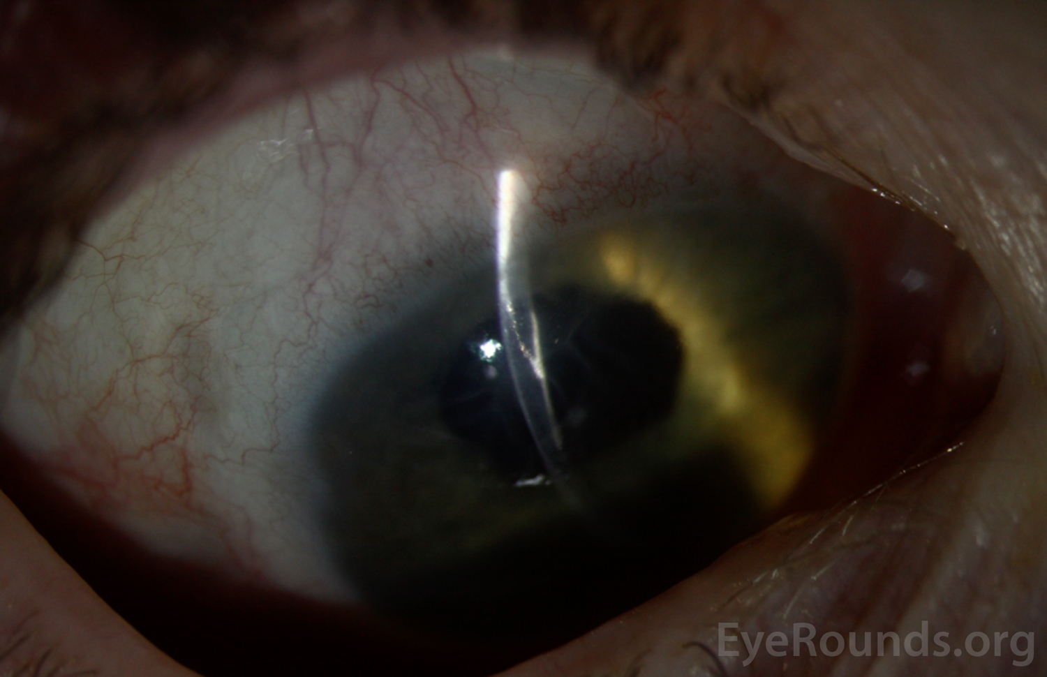
Figure 2: Color slit lamp photograph (narrow beam) of the right eye demonstrating hyperreflective cells extending along the clear corneal incision site.

Dilated fundus examination (DFE)

  • OD
    • Vitreous: Posterior vitreous detachment
    • Disc: Nearly complete cupping, pallor, peripapillary atrophy
    • Cup to disc (C/D): 0.9
    • Macula: Retinal pigment epithelial mottling
    • Vessels: Attenuated
    • Periphery: Grossly attached
  • OS
    • Vitreous: Normal
    • Disc: Mild pallor, peripapillary atrophy
    • Macula: Retinal pigment epithelial mottling
    • Vessels: Attenuated
    • Periphery: Not well visualized

Additional testing

  • Anterior segment optical coherence tomography, shown in Figure 3.

Anterior segment optical coherence tomography (AS-OCT) of the right eye demonstrating a hyperreflective sheet extending through the previous surgical incision and spreading across the endothelium.

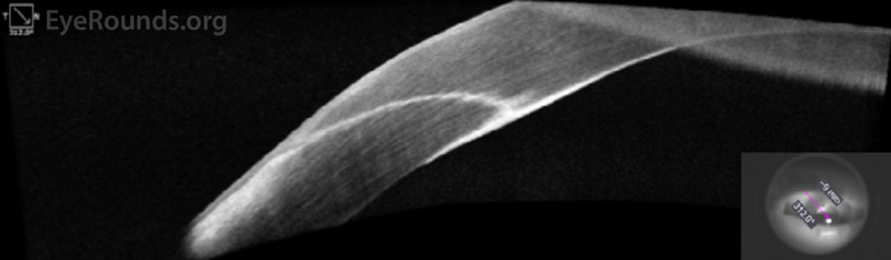
Figure 3: Anterior segment optical coherence tomography (AS-OCT) of the right eye demonstrating a hyperreflective sheet extending through the previous surgical incision and spreading across the endothelium.

Differential Diagnosis

  • Epithelial downgrowth
  • Fibrous downgrowth
  • Pseudophakic bullous keratopathy
  • Fuchs endothelial corneal dystrophy
  • Congenital thick cornea
  • Corneal scar

CLINICAL COURSE

The patient's history of numerous ocular surgeries coupled with the exam findings support a diagnosis of epithelial downgrowth. Management options, including careful surveillance, intracameral 5-fluorouracil (5-FU), cryotherapy, membrane peel, excision, and endothelial keratoplasty, were discussed with the patient.

The degree of blurred vision (20/800) was felt to be out of proportion to the corneal findings. The corneal edema was confined to the superotemporal quadrant and minimally involved the central visual axis. Central pachymetry was thickened but comparable to the contralateral eye. It was thought the patient may have thick corneas at baseline related to his congenital ocular syndrome. Thus, the decision was made to proceed with careful surveillance while continuing Muro128.

DIAGNOSIS

Epithelial downgrowth

DISCUSSION

Epithelial downgrowth is a rare but potentially devastating complication that can occur following any intraocular surgery or penetrating ocular trauma. The proliferation of epithelial cells within the eye can lead to endothelial decompensation, recalcitrant glaucoma, and severe vision loss. Treatment is variable and, unfortunately, often ineffective.

Etiology/Epidemiology

The first reported case of epithelial invasion into the anterior chamber following a perforating eye injury was in 1832. During this era, epithelial downgrowth was cited as the cause of up to a quarter of all enucleations after cataract surgery.[1] Advances in surgical technique, including improved corneal wound construction, have greatly reduced the incidence of this vision-threatening complication.[1 ,2] The most recently cited incidence of epithelial downgrowth following cataract surgery was 0.076 - 0.12% at Massachusetts Eye and Ear Hospital over the years 1953 – 1983.[1] Common risk factors are cataract surgery, penetrating trauma, and penetrating keratoplasty. Epithelial downgrowth most often presents within the first year of the inciting event, but has been reported to occur up to 10 years post-operatively or post-trauma.[3]

Pathophysiology

Epithelial invasion occurs when non-keratinized squamous epithelial cells from the conjunctiva or cornea migrate through a surgical or traumatic corneal wound. This can occur either at the time of surgery or trauma, or secondarily through an incompetent wound or fistula. Additional risk factors include repeated ocular procedures,[4] vascularized corneal stroma, and damaged endothelium, which may predispose to epithelial migration due to loss of normal contact inhibition.[5]
Commonly, the proliferating cells take on the form of a retrocorneal sheet. Other less common presentations include cysts or free-floating cells.

Signs/Symptoms

Patients classically present with painless decrease in vision, but redness, pain, tearing, and photophobia are also common.[1] Slit-lamp exam usually demonstrates a retrocorneal translucent or grey membrane extending from a prior surgical or traumatic wound. Careful gonioscopy and dilated exam should be performed to assess for epithelial growth in the iridocorneal angle or posterior pole, respectively.

The degree of ocular morbidity depends on which structures are affected.  These may include the corneal endothelium (with subsequent corneal edema), iridocorneal angle (leading to obstruction of the trabecular meshwork), iris (with distortion or irregularity of the iris stroma), crystalline lens (cataract) or artificial intraocular lens (sheet), vitreous body (free-floating cells) and retina.[6] With the growing popularity of endothelial keratoplasty (EK) procedures, epithelial cells can also be found within the EK-stroma interface.

Secondary complications include chronic inflammation, corneal decompensation, pupillary block, secondary glaucoma, tractional retinal detachment, phthisis bulbi, and permanent loss of vision.[1 ,7]

Testing and Imaging Work-up

Anterior segment OCT may demonstrate a hyperreflective sheet extending from a previous surgical or traumatic wound. Corneal confocal microscopy will reveal round, nucleated epithelial cells displacing normal hexagonal endothelial cells.[8] If the epithelial sheet involves the iris stroma, laser photocoagulation will confirm the diagnosis by causing the sheet of abnormal cells to blanch,[9 ,10] compared to  laser burns to uninvolved iris which turn dark brown or black. Aqueous aspiration with subsequent cytologic examination can assess for the presence of free-floating epithelial cells.[11]

Treatment/Management/Guidelines 

Proliferating epithelial cells are difficult to remove and often recur despite aggressive treatment. Sheet-like growth, the most common presentation, has the worst prognosis due to difficulty identifying the borders of the lesion and the destructive nature of the surgical procedures required to excise it. In the late 1960s, Maumenee et al found that 27.5% of eyes surgically treated for epithelial downgrowth had vision of 20/50 or better.[12] In a clinicopathological review of 124 patients with epithelial downgrowth from 1953-1983, 50% of patients treated surgically eventually lost vision, required enucleation, or had phthisis bulbi.[1] More recent techniques may lead to improved outcomes.

Treatment depends on the location and extent of involvement. Localized epithelial cells can be devitalized using transcorneal cryotherapy or laser photocoagulation.[13] A confined retrocorneal membrane may be surgically scraped off. However, more extensive involvement may require en-bloc surgical excision of all compromised tissue.[14] In addition, care must be taken not to seed epithelial cells further. For instance, in the case of epithelial cells within an EK graft-stromal interface, complete removal with a penetrating keratoplasty, as opposed to repeat EK, is recommended.[15]

In an effort to reduce surgical destruction of tissue, physicians have started treating epithelial downgrowth with intracameral 5-fluorouracil (5-FU),[3 ,16] which is a  pyrimidine-analog antimetabolite. 5-FU selectively inhibits rapidly dividing cells. It has other ophthalmic applications including topical treatment for ocular surface squamous neoplasia and prevention of fibrosis after trabeculectomies. Various methods have been employed to concentrate delivery of the medication intracamerally for treatment of epithelial downgrowth, including mixing 5-FU with viscoelastic followed by with face-down positioning by the patient.[17]

Management of the cyst form of epithelial downgrowth has a higher success rate. Cystic lesions are easily identifiable and the implanted cells are circumscribed within the cyst. Various techniques have been described, such as aspiration of the cyst, intra-cystic irrigation with a sclerosing agent such as absolute alcohol, and surgical excision of the cyst including its stalk.[18-20]

Whatever the treatment modality chosen, careful surveillance is required to identify any recurrence of these aggressively replicating cells.

EPIDEMIOLOGY OR ETIOLOGY
  • Rare
  • Complication of intraocular surgery or penetrating trauma
  • Intraocular migration of epithelial cells from cornea or conjunctiva
SIGNS
  • Retrocorneal translucent or grey membrane
  • Free-floating or anchored cyst (less common)
  • Anterior chamber inflammation
  • Endothelial decompensation, angle-closure glaucoma, or tractional retinal detachment
SYMPTOMS
  • Decreased vision
  • Pain and photophobia
  • May be asymptomatic
TREATMENT/MANAGEMENT
  • Careful observation
  • Cryotherapy
  • Laser photocoagulation
  • En-bloc surgical excision
  • Intracameral 5-FU injection
References
  1. Weiner MJ, Trentacoste J, Pon DM, Albert DM. Epithelial downgrowth: a 30-year clinicopathological review. Br J Ophthalmol 1989;73(1):6-11. [PMID 2920156]
  2. Vargas LG, Vroman DT, Solomon KD, Holzer MP, Escobar-Gomez M, Schmidbauer JM, Apple DJ. Epithelial downgrowth after clear cornea phacoemulsification: report of two cases and review of the literature. Ophthalmology 2002;109(12):2331-2335. [PMID 12466179]
  3. Kim SK, Ibarra MS, Syed NA, Sulewski ME, Orlin SE. Development of epithelial downgrowth several decades after intraocular surgery. Cornea 2005;24(1):108-109. [PMID 15604876]
  4. Chen SH, Pineda R, 2nd. Epithelial and fibrous downgrowth: mechanisms of disease. Ophthalmol Clin North Am 2002;15(1):41-48. [PMID 12064080]
  5. Davanger M, Olsen EG. Experimental epithelial ingrowth. Epithelial/endothelial interaction through a corneal perforation, studied in organ culture. Acta Ophthalmol (Copenh) 1985;63(4):443-448. [PMID 4050366]
  6. McDonnell JM, Liggett PE, McDonnell PJ. Traction retinal detachment due to preretinal proliferation of surface epithelium. Retina 1992;12(3):248-250. [PMID 1410834]
  7. Patel HR, Margo CE. Epithelial downgrowth. Report of 2 cases diagnosed by ocular biopsy. Ann Diagn Pathol 2017;26:60-63. [PMID 28209236]
  8. Chen MC, Cortes DE, Harocopos G, Mannis MJ. Epithelial downgrowth after penetrating keratoplasty: imaging by high-resolution optical coherence tomography and in vivo confocal microscopy. Cornea 2013;32(11):1505-1508. [PMID 23928949]
  9. Stark WJ, Michels RG, Maumenee AE, Cupples H. Surgical management of epithelial ingrowth. Am J Ophthalmol 1978;85(6):772-780. [PMID 567013]
  10. Lee BL, Gaton DD, Weinreb RN. Epithelial downgrowth following phacoemulsification through a clear cornea. Arch Ophthalmol 1999;117(2):283. [PMID 10037585]
  11. Girkin CA. Glaucoma. Section 10, Basic and Clinical Science Course. San Francisco: American Academy of Ophthalmology, 2018; Chapter 5: Angle-Closure Glaucoma.
  12. Maumenee AE, Paton D, Morse PH, Butner R. Review of 40 histologically proven cases of epithelial downgrowth following cataract extraction and suggested surgical management. Am J Ophthalmol 1970;69(4):598-603. [PMID 4908746]
  13. Han KE, Kim CY, Chung JL, Hong JP, Sgrignoli B, Kim EK. Successful argon laser photocoagulation of diffuse epithelial ingrowth following concomitant persistent pupillary membrane removal and phacoemulsification. J Cataract Refract Surg 2012;38(5):906-911. [PMID 22386278]
  14. Ganesh A, Al-Fadhil N, Wali U, Bialasiewicz AA. En bloc excision of intraocular epithelial cystic downgrowth using syngeneic auricular cartilage. Eye (Lond) 2005;19(1):97-100. [PMID 15286674]
  15. Walker BM, Hindman HB, Ebrahimi KB, Green WR, Eberhart CG, Garcia I, Jun AS. Epithelial downgrowth following Descemet's-stripping automated endothelial keratoplasty. Arch Ophthalmol 2008;126(2):278-280. [PMID 18268229]
  16. Jabbour S, Talajic J, Mabon M, Modabber M, Choremis J. Descemet membrane endothelial keratoplasty for epithelial downgrowth after cataract surgery. Can J Ophthalmol 2019;54(3):e151-e153. [PMID 31109506]
  17. Shaikh AA, Damji KF, Mintsioulis G, Gupta SK, Kertes PJ. Bilateral epithelial downgrowth managed in one eye with intraocular 5-fluorouracil. Arch Ophthalmol 2002;120(10):1396-1398. [PMID 12365927]
  18. Shields CL, Arepalli S, Lally EB, Lally SE, Shields JA. Iris stromal cyst management with absolute alcohol-induced sclerosis in 16 patients. JAMA Ophthalmol 2014;132(6):703-708. [PMID 24723076]
  19. Behrouzi Z, Khodadoust A. Epithelial iris cyst treatment with intracystic ethanol irrigation. Ophthalmology 2003;110(8):1601-1605. [PMID 12917180]
  20. Gokhale NS. Management of recurrent intra corneal epithelial cyst with ethanol irrigation and vacuum-assisted cyst wall excision. Int Ophthalmol 2013;33(4):429-433. [PMID 23160824]
Suggested citation format:

Colwell L, Diel RJ, Benage M, Jiang L, Greiner MA, Ling JJ. Epithelial Downgrowth. EyeRounds.org. Posted February 24, 2020; Available from https://EyeRounds.org/cases/292-epithelial-downgrowth.htm


last updated: 2/26/2020

Image Permissions:
Creative Commons

Ophthalmic Atlas Images by EyeRounds.org, The University of Iowa are licensed under a Creative Commons Attribution-NonCommercial-NoDerivs 3.0 Unported License.