
INITIAL PRESENTATION
Chief Complaint: Bilateral eye pain
History of Present Illness: A 17-year-old boy presented with one day of acute-onset bilateral foreign body sensation, tearing, photophobia, blurriness, and redness. His symptoms were worse at the end of the day. He denied ocular itching, discharge, contact lens use, known foreign matter exposure, history of upper respiratory illness, sick contacts, and cold sores. One week prior to presentation, he was admitted to the hospital for treatment of acute myeloid leukemia (AML). His chemotherapy regimen included cytarabine, which he had received four days prior to the onset of symptoms.
Past Ocular History: Myopia
Medical History: Acute myeloid leukemia (AML) with pancytopenia
Medications:
Allergies: None
Family History: Non-contributory
Social History:
Review of Systems: Positive for fatigue, weight loss, pallor, and petechiae. Otherwise unremarkable other than as noted in history of present illness.
OCULAR EXAMINATION
Full and orthotropic in both eyes (OU)
Full OU
Normal OU
|
OD |
OS |
|
|
Lids/lashes |
Normal |
Normal |
|
Conjunctiva/sclera |
1+ injection without chemosis, follicles, or papillae |
1+ injection without chemosis, follicles, or papillae |
|
Cornea |
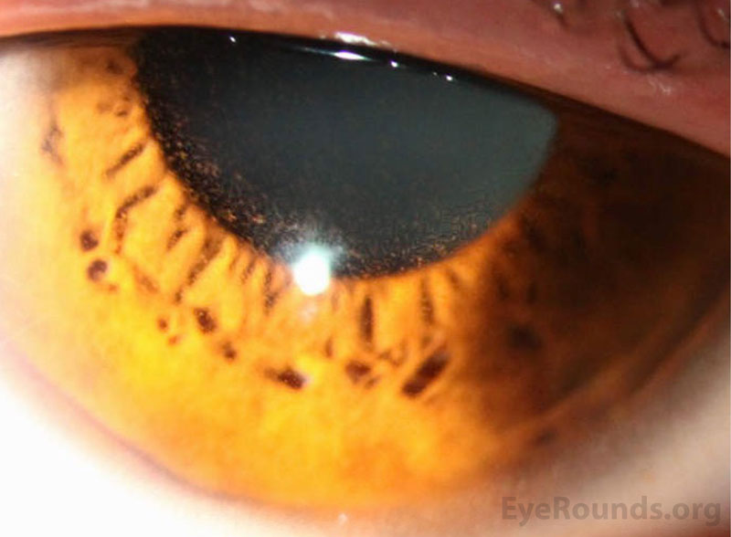
Tiny intraepithelial microcysts scattered diffusely and 1+ punctate epithelial erosions scattered diffusely |
Tiny intraepithelial microcysts scattered diffusely and 1+ punctate epithelial erosions scattered diffusely |
|
Anterior chamber |
Deep & quiet |
Deep & quiet |
|
Iris |
Normal |
Normal |
|
Lens |
Clear |
Clear |
|
Anterior vitreous |
Normal |
Normal |
|
OD |
OS |
|
|
Vitreous |
Clear |
Clear |
|
Disc |
Normal |
Normal |
|
Cup-to-disc ratio: |
0.3 |
0.3 |
|
Macula |
Normal |
Normal |
|
Vessels |
Normal |
Normal |
|
Periphery |
Normal |
Normal |
Differential diagnosis:
CLINICAL COURSE
Given the onset of his symptoms shortly after starting chemotherapy and the lack of evidence of infectious etiology, the patient was diagnosed with cytarabine-induced keratoconjunctivitis. Prednisolone acetate 1% four times daily and artificial tears four times daily were begun. Given his immunosuppressed state, the presence of punctate epithelial erosions, and the initiation of topical steroids, he was also started on Polytrim three times daily to prevent a secondary bacterial infection.
One-week later, his ocular symptoms had resolved, and his intraepithelial cysts were improved. His topical steroid was accordingly tapered off over one month. At his one-month follow-up, he continued to be asymptomatic and his corneas appeared normal.
DIAGNOSIS: Cytarabine-induced keratoconjunctivitis
DISCUSSION
Introduction
Simultaneous corneal and conjunctival inflammation, or keratoconjunctivitis, is a well-described side effect of high-dose systemic cytarabine.[1,2] Cytarabine is an antineoplastic agent commonly used in the treatment of AML and other hematologic malignancies.[3] Routinely, patients beginning high-dose cytarabine are prescribed prophylactic corticosteroid eye drops for the prevention of ocular toxicity.[4]
Epidemiology
Cytarabine-induced keratoconjunctivitis occurs in 40-100% of patients receiving high-dose therapy. The likelihood of developing this side effect is directly associated with the treatment dose and duration.[4-6] This incidence lowers significantly to 8-16% in patients receiving prophylactic corticosteroid eye drops.[4,7]
Etiology and Pathophysiology
Cytarabine is a powerful anti-metabolite that works by inhibiting DNA synthesis. It is a common chemotherapeutic agent in the treatment of malignancies [2]. It can cross the blood-brain barrier, penetrate bodily fluids, and has been found in ocular fluids including the tears and aqueous humor. [8,9,10] Cytarabine-induced keratoconjunctivitis likely develops secondary to inhibition of DNA synthesis in the rapidly dividing corneal and conjunctival epithelium. Specifically, it is thought that the more differentiated and rapidly dividing transient amplifying cells (TACs) located within the corneal epithelial basal layer are more vulnerable to the cytotoxicity of cytarabine than the corneal epithelial stem cells, which have a longer cell cycle duration.[8] Further, TACs are more densely populated in the central cornea, whereas corneal epithelial stem cells predominate peripherally.[8] This may explain the central predominance of the epithelial microcysts in cases of cytarabine-induced keratoconjunctivitis.[8]
Resultant corneal toxicity is proportional to the duration of drug exposure as well as the concentration of cytarabine in the tear fluid.[9,10] As mentioned previously, the risk of developing cytarabine-induced keratoconjunctivitis increases with dose (>1000 mg/m2 body surface area), duration of exposure (>5 days), and concurrent total body irradiation.[6,8,11] However, toxicity has been observed with doses as low as 400 mg/m² body surface area daily.[8]
Symptoms and Signs
Presenting symptoms include eye pain, specifically, intense burning or foreign body sensation.[2,5,12] Additional symptoms include photophobia, blurry vision, tearing, and eye redness.[2,5,12] Onset of symptoms usually occurs within several days of completing a 7-day course of cytarabine, but may also occur sooner, as was seen in this case.[6,12]
Slit lamp examination characteristically reveals bilateral tiny corneal refractile intraepithelial microcysts, concentrated more centrally than peripherally.[2] Some cases reveal fine stromal opacities, but the remainder of the cornea exam is typically normal.[13] Other findings include conjunctival injection, chemosis, and blepharospasm.[2] Inflammation usually spares the intraocular space, though few case reports demonstrate associated symmetric anterior uveitis as a complication of cytarabine treatment.[14-17]
Histologic specimens from patients with cytarabine-induced keratoconjunctivitis reveal replacement of basal columnar cells of the cornea with flattened cells, indicating damage to the cells in this layer.[2] Electron microscopy findings are also consistent with DNA damage to the corneal epithelial cells.[2]
Treatment and Management
Mainstay therapy includes topical corticosteroids and artificial tears for treatment of the ocular surface.[6,8,18,19] Topical steroids may work by decreasing the rate of DNA replication in corneal epithelial cells so that cells in rapid cell cycles (i.e. TACs within the basal level) are less vulnerable to damage.[20] Typical regimens may include prednisolone acetate 1%, dexamethasone 0.1%, and betamethasone 0.1% ranging from every 2 hours or four times daily. Severe cases may necessitate adding topical non-steroidal inflammatory agents such as diclofenac sodium 0.1% every 8 hours.[8,18] Aggressive lubrication with preservative-free artificial tears is vital for symptom control and may also dilute the concentration of cytarabine in tears to reduce inflammation.[6]
Prophylaxis with corticosteroid drops is typically recommended for doses >1000 mg/m2 body surface area and in any patient with prior history of cytarabine-induced keratoconjunctivitis.[7] Typical regimens include prednisolone acetate 1%, dexamethasone 0.1%, and betamethasone 0.1% ranging from every 4 to 6 hours.[6]
Prognosis and Complications
Cytarabine-induced keratoconjunctivitis is generally self-limited with reversible corneal damage. Symptoms generally resolve within a week of therapy initiation. However, intraepithelial microcysts may be seen up to 4 weeks post-treatment.[2]
Summary
Cytarabine-induced keratoconjunctivitis is a common side effect of high-dose therapy used in the treatment of AML and other hematologic malignancies.[1-3] Symptoms include ocular pain, photophobia, blurry vision, tearing, and eye redness.[2,5,12] Slit lamp examination characteristically reveals tiny corneal intraepithelial microcysts, fine corneal opacities, conjunctival injection, chemosis, and blepharospasm.[2,13] When keratoconjunctivitis occurs, mainstay therapy includes topical corticosteroids and artificial tears.[6,8,18,19] These measures, when used prophylactically, have been shown to decrease the incidence of cytarabine-induced keratoconjunctivitis from 40-100% to 8-16%, and is recommended for all patients receiving high-dose cytarabine.[4-7]
EPIDEMIOLOGY AND ETIOLOGY [4-7]
|
SIGNS [2,5,12]
|
SYMPTOMS [2,5,12]
|
MANAGEMENT [6,8,18,19]
|
References
Hendricks T, Quist TS, Ling JL, Greiner MA. Cytarabine-induced Keratoconjunctivitis. EyeRounds.org. January 26, 2023. Available from https://eyerounds.org/cases/338-cytarabine-induced-keratoconjunctivitis.htm

Ophthalmic Atlas Images by EyeRounds.org, The University of Iowa are licensed under a Creative Commons Attribution-NonCommercial-NoDerivs 3.0 Unported License.

Address
University of IowaLegal
Related Links
