This site uses tracking information. Visit our privacy policy. Click to agree to this policy and not see this again.

Ophthalmology and Visual Sciences

Conjunctival intraepithelial neoplasia (CIN) with full thickness dysplasia (squamous cell carcinoma in situ)

Conjunctival intraepithelial neoplasia (CIN) with full thickness dysplasia (squamous cell carcinoma in situ)

Category(ies): Cornea, External Disease
Contributor: John Chen, MD, PhD
Photographer: Cindy Montague, CRA

Conjunctival intraepithelial neoplasia
Conjunctival intraepithelial neoplasia (CIN) with full thickness dysplasia (squamous cell carcinoma in situ)
enlarged Conjunctival intraepithelial neoplasia
Enlarged: Conjunctival intraepithelial neoplasia (CIN) with full thickness dysplasia (squamous cell carcinoma in situ)

Conjunctival Squamous Cell Carcinoma (SCC) in situ

Conjunctival Squamous Cell Carcinoma (SCC) in situ

Category(ies): Cornea / External Eye Disease
Contributor: Ella J. Gehrke, MD; Kanwal Singh Matharu, MD
Photographer: Sarah L. Skiles
Posted: 04/17/2025

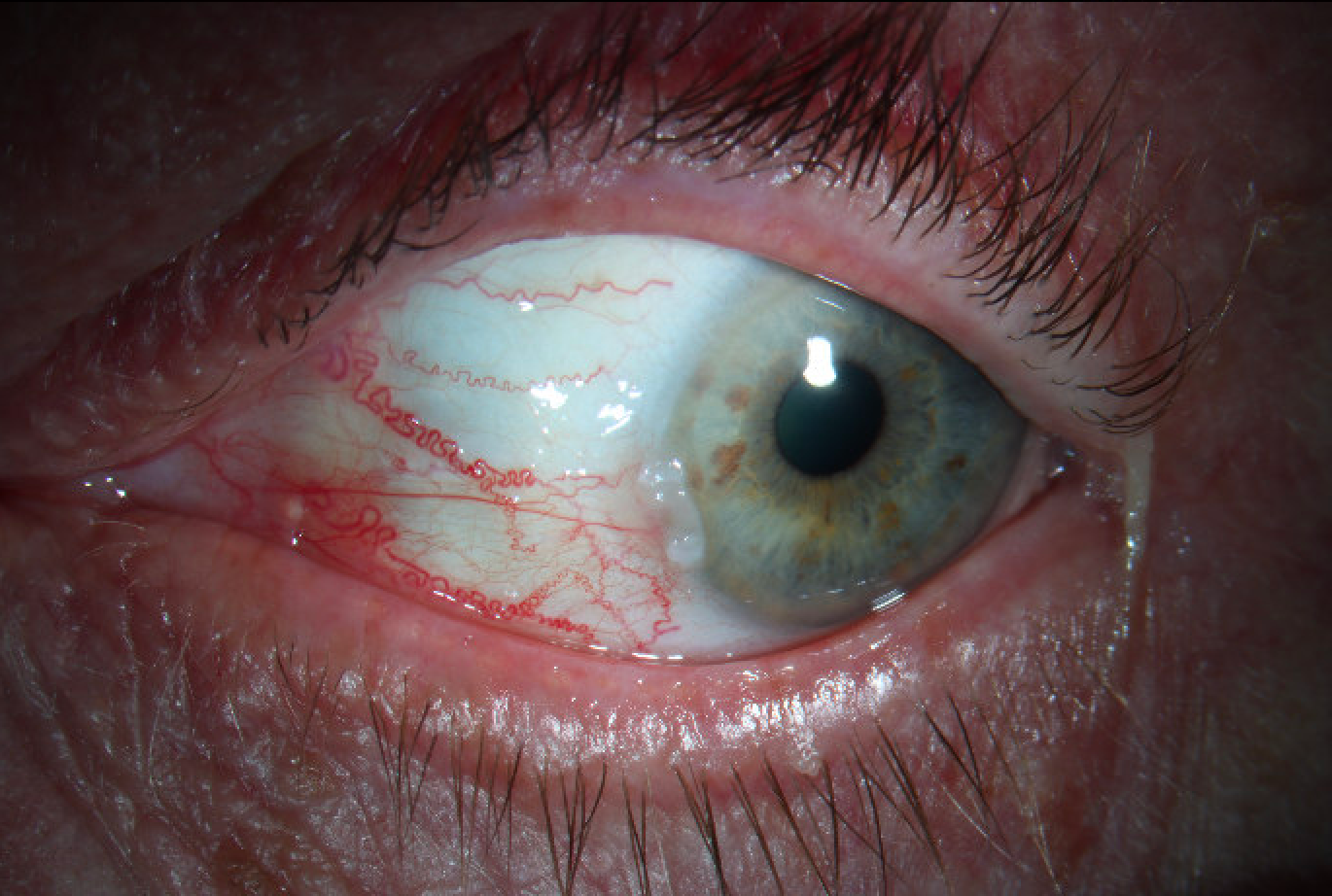
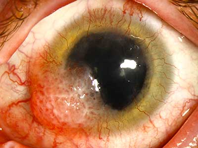
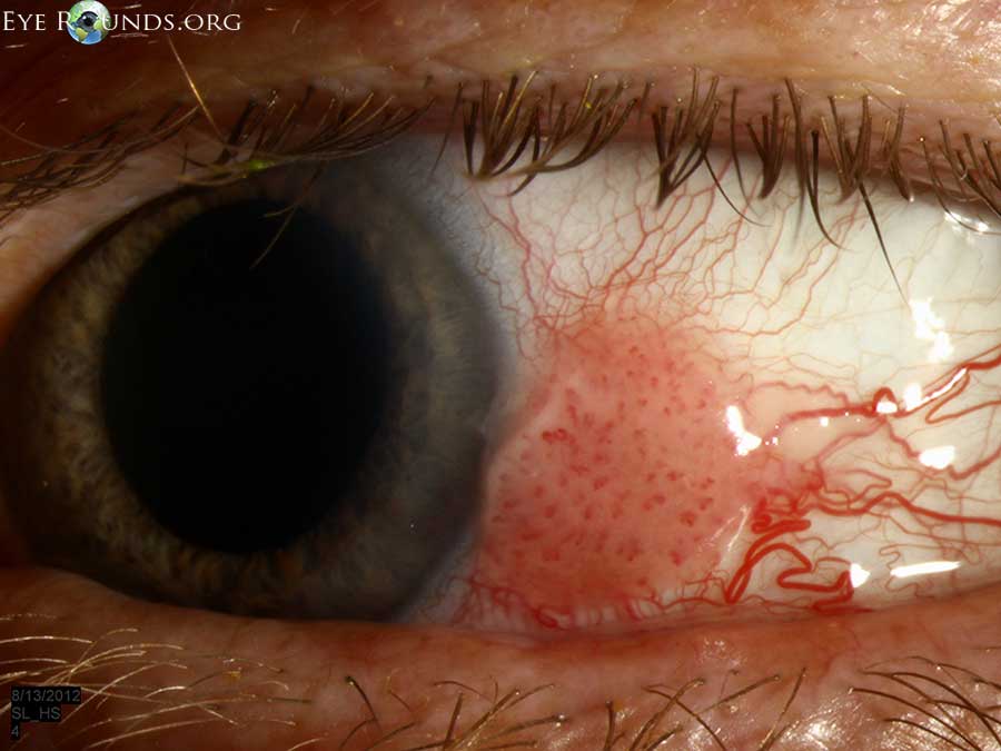
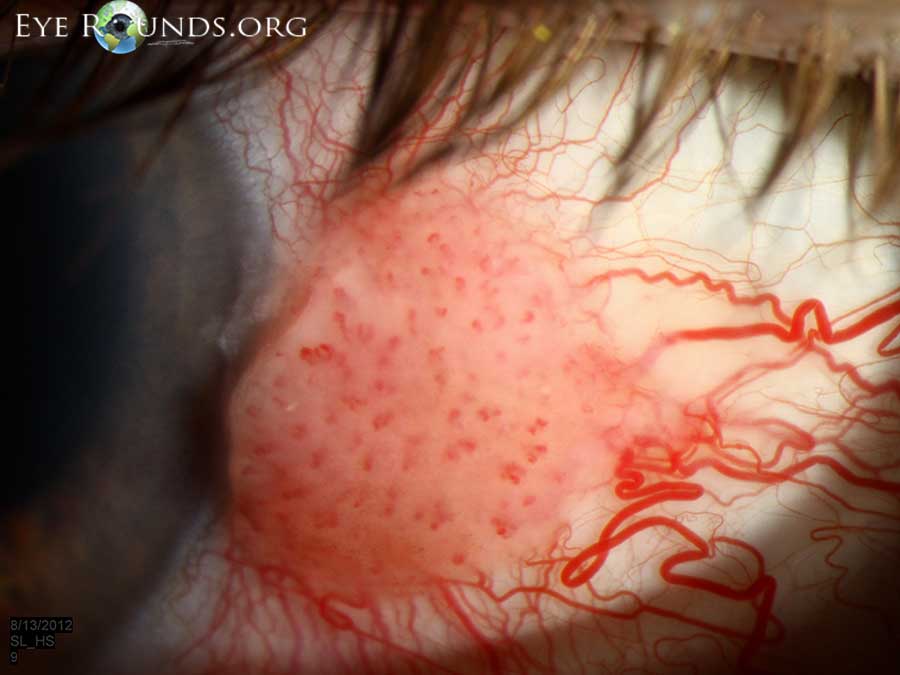
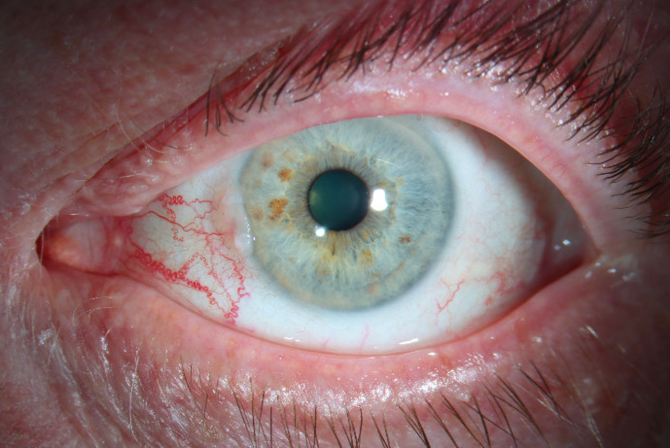
A 68-year-old male patient presented for a routine cataract evaluation and was found to have a suspicious conjunctival lesion in the left eye. He reports symptoms of foreign body sensation, itching, and extreme ocular dryness (notably frequent blinking). The patient has a known history of cryotherapy for precancerous skin lesions on the face. On the exam, the best-corrected visual acuity was 20/20 in both eyes with intraocular pressures of 19 mmHg OD and 20 mmHg OS. Extraocular movements and visual fields were full bilaterally. Slit lamp exam of the left eye revealed a nasal, elevated, gelatinous conjunctival lesion with leukoplakic areas extending approximately 1 mm onto the cornea, along with a temporal pinguecula and prominent nasal vessels. The patient underwent conjunctival biopsy of the left eye, and histopathology revealed a moderately differentiated squamous cell carcinoma in situ with positive margins. The patient underwent 6 rounds of topical chemotherapy therapy with Fluorouracil (5-FU).

Slit lamp photograph of the left eye demonstrates a nasal, elevated, gelatinous, flesh-colored conjunctival lesion with leukoplakic areas extending approximately 1 mm onto the cornea, along with prominent nasal vessels.
Slit lamp photograph of the left eye demonstrates a nasal, elevated, gelatinous conjunctival lesion with leukoplakic areas extending approximately 1 mm onto the cornea, along with prominent nasal vessels.
Slit lamp photograph of the left eye in lateral gaze demonstrates a nasal, elevated, gelatinous, flesh-colored conjunctival lesion with leukoplakic areas extending approximately 1 mm onto the cornea, along with prominent nasal vessels.