
Chief Complaint: Eye crossing
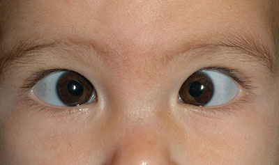
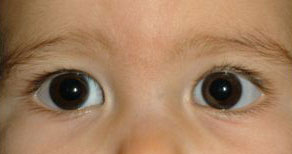
History of Present Illness: The patient is a 2 month-old girl referred by her pediatrician for evaluation of esotropia. Her parents noted that the patient began crossing her eyes shortly after birth and feel that it is worsening. They also note that the crossing is a little worse when she is tired and alternates between the right eye and the left eye.
Medical History: The patient was born full term by normal spontaneous vaginal delivery without complications. Thus far she is a healthy baby, growing and developing normally.
Past Surgical History: None
Family History: No known strabismus or inherited eye disease
Ocular Examination:
Visual Acuity (without correction):
Pupils: Equally round and briskly reactive. No relative afferent pupillary defect.
Stereo Vision: Unable to test
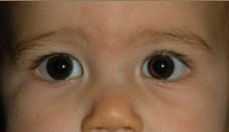
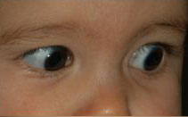
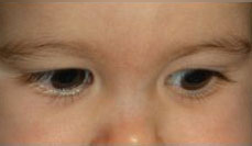
Motility and Strabismus: Krimsky and Hirschberg methods
No palpebral fissure narrowing or globe retraction on evaluation of versions.
Cycloplegic Refraction:
General: Normal mood and behavior; no nystagmus or abnormal head position
External Exam: Epicanthal folds OU
Slit Lamp Exam: Normal anterior segment exam OU without evidence of cataracts or other media opacities. Normal-appearing optic nerves with cup-to-disc ratios of 0.1 OU. Normal fundus examination.
Visual Acuity (without correction)
Motility and Strabismus: Krimsky and Hirschberg methods
Cycloplegic Refraction:
Visual Acuity (without correction)
Motility and Strabismus: Krimsky and Hirschberg methods
 |
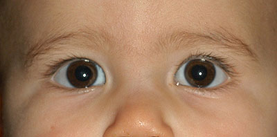
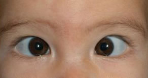
At 6 months of age, she underwent uncomplicated bilateral medial rectus recessions (11.5 mm from the limbus).
Visual Acuity (without correction)
Pupils: Equally round and briskly reactive. No relative afferent pupillary defect. No nystagmus.
Motility and Strabismus: Krimsky and Hirschberg methods
 |
|
 |
|
 |
 |
 |
|
 |
Visual Acuity (without correction)
Motility and Strabismus: by Alternate Cover Testing
 |
 |
| Pre-operative | Post-operative |
The term congenital esotropia is often used interchangeably with infantile esotropia, but few cases are actually noted at birth. Often the exact date of the deviation is not precisely established, but the appearance of an esodeviation by 6 months is widely accepted by ophthalmologists as necessary to make the diagnosis. Some prefer to use the term infantile esotropia to more accurately reflect the typical postnatal onset of misalignment (Nelson, 1987).
Although the exact etiology of infantile esotropia remains unknown, two conflicting theories have been proposed. The first was proposed by Claud Worth in 1903. He suggested that this form of esotropia is "a congenital defect of the fusion faculty" (Worth, 1903). He went on to describe it as an irreversible defect in the brain's ability to fuse, rendering the goal of restoring binocularity hopeless. Worth's "sensory" theory was countered by Chavasse's "motor" theory. In 1939, F. Bernard Chavasse proposed that the neural components of binocular vision are present in congenital esotropia. He reasoned that the primary problem is mechanical and potentially curable if the deviation could be eliminated in infancy. His theory suggests that abnormalities in binocular vision may be the result and not the cause of early strabismus (Chavasse, 1939). Until the 1960's studies seemed to support Worth's view of irreversible loss of binocularity, but over the past fifty years studies have reported favorable sensory results in some infants who underwent surgical correction between 6 months and 2 years. These more recent findings formed the basis for the current practice of early surgery for patients with congenital esotropia.
All children with new-onset esotropia require a thorough history, review of systems, and comprehensive examination. The examination must include assessment of vision, ocular motility, strabismus measurements at distance and near and cycloplegic refraction in addition to general ocular examination. Besides having strabismus, the majority of children with congenital esotropia are developmentally normal. Esotropia, however, can be observed in the setting of neurologic or developmental disorders which highlights the importance of a careful and complete history and exam.
The key to a successful ocular examination is efficient and accurate methods of testing vision, motility, and ocular alignment. One method of visual acuity testing in preverbal or nonverbal children is the CSM method. C refers to centrality and can be assessed by the location of the corneal light reflex as the patient looks at the examiner's light with the opposite eye occluded. Normally, the light reflex should be located near the center of the cornea. S refers to the steadiness of fixation on the examiner's light under monocular conditions. M refers to the ability of the patient to maintain alignment. The ability of an eye to maintain fixation is assessed by occluding the contralateral eye to stimulate fixation on a target and then assessing whether fixation is maintained under binocular conditions when the contralateral eye is unoccluded. If, as the contralateral eye is uncovered, the eye under examination shifts within 3 seconds or before the patient blinks, the examined eye is said to be "unmaintained". The inability to maintain fixation is indicative of amblyopia.
In young children, it can be more challenging to obtain ocular alignment measurements. Since there is limited cooperation for cover testing, estimates of the deviation can be made using corneal light reflex tests. The Hirschberg method is based on the premise that 1 mm of decentration of the corneal light reflex corresponds to approximately 15 prism diopters of ocular deviation. The Krimsky method uses reflection of light produced simultaneously on both corneas. The original method involved placing prism in front of the deviating eye until the corneal reflexes were similar. The modified-Krimsky method involves holding prisms before the fixating eye or splitting prism between the two eyes. Again, as the corneal reflections become similar, a quantifiable estimate is made.
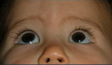
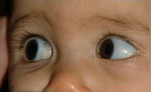
Patients with congenital esotropia often cross-fixate. Cross-fixation occurs when the patient prefers to fixate with either eye in adduction. A pattern of cross-fixation does not infer spontaneous or equal alternation or the absence of amblyopia. Close attention needs to be paid to the point at which the alternation occurs. If amblyopia is present with cross-fixation the alternation will not take place at midline; the sound eye will maintain fixation into abduction. If a child has equal vision and cross-fixates, they have no need to abduct either eye and the examiner will note alternation at midline. This apparent abduction deficit can erroneously lead one to suspect a congenital sixth nerve palsy. Assessing motility and the child's ability to abduct the eye can be demonstrated with the "doll's head" maneuver—also known as vestibular rotation—or by observing motility with one eye occluded (ductions).
This case is an example of a good post-operative outcome with early surgical treatment. The patient presented with a large angle esotropia noted within two months of life. No amblyopia was detected preoperatively. Motility was full and there was no globe retraction—effectively ruling out congenital sixth nerve palsy and Duanes retraction syndrome. A low-normal amount of hyperopia was found on cycloplegic retinoscopy without significant astigmatism or anisometropia, so no glasses were initiated. Should any significant refractive errors be detected, the full cycloplegic refraction should be prescribed. Inferior oblique muscle overaction and dissociated vertical deviation (DVD) occur in more than half of patients with congenital esotropia. Both of these associations are usually not recognized until after 1 year of age.
The primary goal of treating infantile esotropia is to reduce the deviation to orthotropia or as close to orthotropia as possible. In addition to improving alignment of the eyes, additional goals are the development of some degree of sensory fusion and preservation of normal sight in each eye. Alignment within 10 prism diopters of orthotropia often results in the development of a monofixation syndrome. Monofixation syndrome is characterized by peripheral fusion and central suppression. Even though a macular scotoma develops in the non-fixating eye (central suppression), binocular fusion of points in the peripheral retina is retained (peripheral fusion). This small angle deviation generally represents a stable, functional surgical outcome, produces a favorable cosmetic appearance and is considered a surgical success even though bifoveal fusion is not achieved.
Various surgical approaches have been described for the management of congenital esotropia. The most common procedure is bilateral medial rectus recession. An acceptable alternative is a recess/resect procedure or "R&R," which is recession of a medial rectus muscle combined with resection of the ipsilateral lateral rectus muscle. Two-muscle surgery spares the remaining two horizontal muscles for subsequent surgery should it be needed. Studies vary widely in estimates of re-operation but a moderate portion of patients will require an additional surgery in their lifetime. Some surgeons will perform initial 3- or 4-muscle surgeries if the primary deviation is greater than 50 prism diopters (Scott, 1986).
It is important for the ophthalmologist to take time to discuss the condition with the parents or caretakers. It is also important to convey that this is a long-term condition that requires continued monitoring for amblyopia and stability in ocular alignment. It should be mentioned that a moderate number of patients will require additional surgery at some point in their lives to achieve adequate and stable alignment. Also, latent nystagmus occurs in up to one third of patients with a history of early-onset esotropia (Hiles, 1980). And as mentioned earlier, inferior oblique muscle overaction and dissociated vertical deviation (DVD) occur in more than half of patients with congenital esotropia. Parents are rightfully concerned over their child's long term prognosis, and discussion regarding these apprehensions should be addressed.
Summary
|
Signs
|
Symptoms
|
Treatment
|
Nelson LB, Wagner RS, Simon JW, et al. Congenital esotropia. Surv ophthalmol 1987; 31(6): 363-383.
Worth CA. Squint. Its Causes, Pathology, and Treatment. Philadelphia & London: Blakiston, 1903; p. 4.
Cavasse FB. Worth's squint or the binocular reflexes and the treatment of strabismus. London: Baillier, Tindall and Cox, 1939; p. 519.
Scott WE, Reese PD, Hirsch Cr, et al. Surgery for large angle congenital esotropia. Arch ophthalmol 1986; 104:374-7.
Hiles DA, Watson A, Biglan AW. Characteristics of infantile esotropia following early bimedial rectus recession. Arch ophthalmol 1980; 98:697-703.
Rogers GM, Pfeifer WL, Longmuir SQ. Infantile (Congenital) Esotropia. Eyerounds.org. November 6, 2010; Available from: http://www.EyeRounds.org/cases/124-infantile-esotropia.htm

Ophthalmic Atlas Images by EyeRounds.org, The University of Iowa are licensed under a Creative Commons Attribution-NonCommercial-NoDerivs 3.0 Unported License.

Address
University of IowaLegal
Related Links
