This site uses tracking information. Visit our privacy policy. Click to agree to this policy and not see this again.

Ophthalmology and Visual Sciences

Phacolytic glaucoma in an eye with no light perception

Dislocated dense cataract inducing inflammation in an eye with no light perception

Phacolytic glaucoma in an eye with no light perception

Dislocated dense cataract inducing inflammation in an eye with no light perception
Hend M. Al-Kaylani, BS; Eric A. Lovett Jr., MBA, BS; Adetayo Oladele-Ajose, MD; Erin Boese, MD
February 24, 2026

INITIAL PRESENTATION

Chief Complaint: Ache in the left eye

History of Present Illness:

An 86-year-old male presented to clinic with one week of pain in the left eye. The pain was described as a constant, aching, pressure-like sensation that radiated posteriorly and was rated 4/10. Pain worsened with eye movement, especially with left or right gaze. He tried acetaminophen and antihistamine drops, which did not help. He reported closing his eyes alleviated some of the pain temporarily. He also experienced a foreign body sensation, redness, and tearing of the left eye. Notably, he had a history of trauma to the left eye, leaving him no-light-perception (NLP) over 60 years ago. His right eye remained asymptomatic. He denied chills, malaise, new aches, or fatigue.

Past Ocular History:

  • Retained metallic intraocular foreign body OS, secondary to trauma 63 years prior
    • Wears polycarbonate lenses for monocular precautions
  • Cataract extraction with intraocular lens placement OD
  • Dense brunescent cataract OS with dislocation posteriorly.
  • Non-proliferative diabetic retinopathy, mild, with clinically significant diabetic macular edema OD
    • Treated with aflibercept 2x 8 years ago, observed since then
  • Glaucoma suspect
    • Timolol daily OD
  • Dry eye OU
    • Artificial tears TID, warm compresses BID

Past Medical History:

  • Type 2 Diabetes Mellitus, insulin-dependent
  • Hypertension
  • Heart failure with reduced ejection fraction
  • Atrial fibrillation
  • Chronic kidney disease stage 3b

Medications:

  • Semaglutide 0.5 mg SQ weekly
  • Basal-bolus insulin
  • Apixaban 5 mg BID
  • Metoprolol 12.5 mg BID
  • Fludrocortisone 0.05 mg daily

Allergies:

  • No known allergies

Family History:

  • Non-contributory

Social History:

  • Lives in a nursing home

Review of Systems:

  • Negative except for what is detailed in the history of present illness.

OCULAR EXAMINATION

  • Visual Acuity without correction (on Snellen chart):
    • Right eye (OD): 20/25+2, no improvement with pinhole
    • Left eye (OS): NLP
  • Ocular Motility/Alignment:
    • Full OU
  • Intraocular Pressure (IOP) (Tonopen):
    • OD: 13 mmHg
    • OS: 9 mmHg
  • External:
    • Unremarkable OU
  • Slit Lamp Examination:
    • Lids/Lashes: Meibomian gland dysfunction OU
    • Conjunctiva/sclera:
      • Clear, quiet OD
      • Trace injection, nasal pterygium 2 mm onto cornea OS
    • Cornea:
      • Inferior punctate epithelial erosions OD
      • Peripheral clouding OS
    • Anterior chamber:
      • Deep and quiet OD
      • 3-4+ cell, 1-2+ flare OS
    • Iris:
      • Pharmacologically dilated OD
      • Transillumination defect at 7:00 OS
    • Lens:
      • PCIOL, centered OD
      • Aphakia OS
    • Anterior vitreous
      • Quiet OD
      • 3+ Cell
  • Dilated Fundus Examination (DFE):
    • Vitreous:
      • Posterior vitreous detachment OD
    • Disc:
      • Pink, sharp border OD
      • No disc hemorrhage OU
    • Cup-to-disc ratio:
      • Large disc, 0.8 OU
    • Macula:
      • Microaneurysms OU
    • Vessels:
      • Normal OU
    • Periphery:
      • Microaneurysms OU
      • Large inferior dome-shaped mass near area of atrophy OS
  • This is a fundus photo of the left eye showing clear media, diffusely pallid disc, a chorioretinal scar along the inferotemporal arcades near the equator, and a large bulbous mass in the inferior quadrant.
    Enlarge Download
    Figure 1. This is a fundus photo of the left eye showing clear media, diffusely pallid disc, a chorioretinal scar along the inferotemporal arcades near the equator, and a large bulbous mass in the inferior quadrant.
  • Additional Testing:
    • B-scan ocular ultrasound was obtained in the left eye on initial presentation; findings are presented in Figures 2a, 2b (photos) and Figure 3 (video).
    Figure 2a,2b. This is a formal B-scan oriented at L3 (2a, top) and T6E (2b, bottom), which shows the dense cataractous dislocated lens has fallen to the back and is resting on the retina. Notice the completely intact lens capsule.
  • Fluorescein angiography of the left eye at 47 seconds showing nonperfusion from BRAO within the superior aspect of the macula with multiple foci of segmental arterial wall hyperfluorescence superior to the disc and along the inferior arcade.
    Enlarge Download
    Figure 3. A hyperechoic, biconvex mass is identified in the posterior chamber using B-scan ocular ultrasound.
    On slit lamp exam, Cell and flare is visible in the left anterior chamber, and the lens is not identifiable in the posterior chamber.
    Enlarge Download
    Figure 4. On slit lamp exam, Cell and flare is visible in the left anterior chamber, and the lens is not identifiable in the posterior chamber.

DIFFERENTIAL DIAGNOSIS:


CLINICAL COURSE

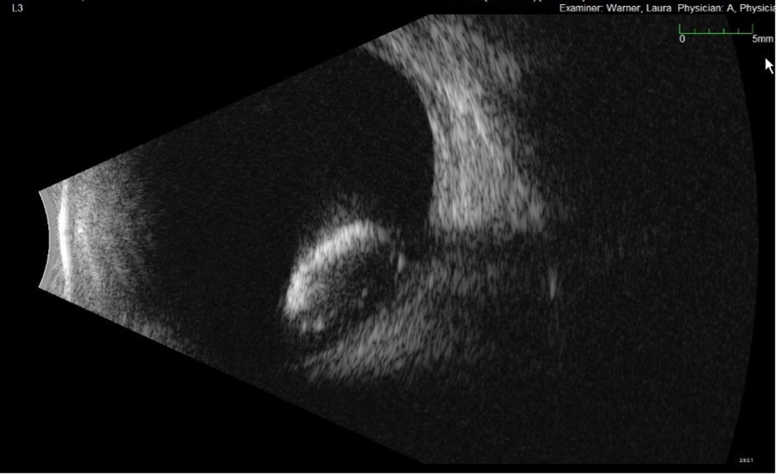
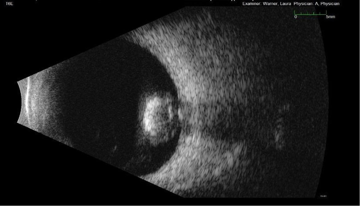
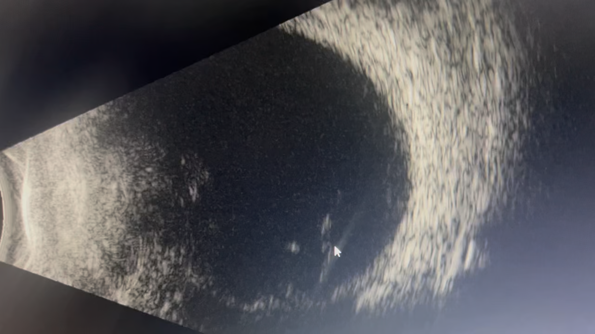
The patient was initially treated for possible stroke vs MS flare at the outside hosUltrasound of the orbit showed a lens material dislocated into the posterior segment with vitreous debris leaking protein, consistent with phacolytic glaucoma. The patient was placed on prednisolone 1% four times daily. Given the eye had no light perception at baseline, vitrectomy and/or cataract extraction were not indicated.

One week after prednisolone therapy, the pain resolved. Slit lamp exam revealed 1+ cell with flare, improved from 2-3+ cell and flare on initial presentation. Intraocular pressure was 14 mmHg. Risk for sympathetic ophthalmia was determined to be low given that the eye/ lens capsule had not been violated. Utilizing shared decision-making with the patient, we decided to continue watchful waiting given that the eye was painless. We discussed red flag signs and symptoms for pressure spikes in case the patient had a steroid response. We were less concerned for asymptomatic high pressures given that this was an NLP eye. We also broached the topic of enucleation in the future if the eye became painful.

The patient was seen again at week 3 and found to have complete resolution of all anterior chamber and anterior vitreous inflammation and pressure had elevated to 23 mmHg, but the patient denied any pain. The patient was continued on prednisolone 1% four times daily with a plan to taper if he starts to have symptomatically elevated pressures, and he will return in 6 months for check in.


DIAGNOSIS: Phacolytic glaucoma


DISCUSSION

Etiology/Epidemiology:

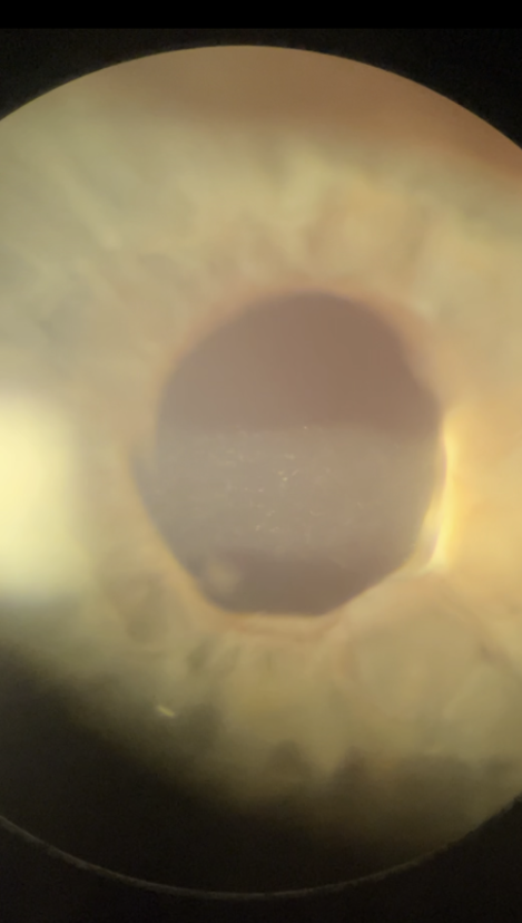
Phacolytic glaucoma is a secondary open-angle glaucoma in which leaked lens proteins from a hypermature cataract induce an inflammatory reaction in the anterior chamber, disrupting aqueous humor outflow(1). Most patients are over the age of 60, given age is the strongest risk factor for hypermature cataract formation. There is a slightly higher predilection in females, with one study citing a 1.7:1 female to male in incidence. The diagnosis is more commonly seen in patients from resource-limited areas with barriers to healthcare or who have a contraindication to cataract extraction in the eye (ex. no light perception in our patient)(2).

Signs/Symptoms:

It is important to recognize that vision loss will likely not be the patient’s chief concern, given a hypermature cataract will have established chronic vision loss prior to any inflammatory reaction(3). Thus, most common chief concerns include pain, epiphora, or conjunctival injection. On slit lamp examination, lens cortical particulates may be visible in the anterior chamber. Signs that distinguish phacolytic from phacoantigenic glaucoma include the notable lack of keratic precipitates in phacolytic glaucoma(4). While increased intraocular pressure is a feature of most cases, it is not required. In our case, preexisting damage to the ciliary body resulted in comorbid reduced aqueous humor production and a low intraocular pressure despite the disruption in aqueous humor outflow.

Testing/Laboratory work-up:

Anterior chamber paracentesis can be performed to decrease intraocular pressure and collect fluid for cytopathology. Light microscopy shows activated epithelioid histiocytes containing lenticular material, but an absence of histiocytes does not rule out the diagnosis(5).

Imaging:

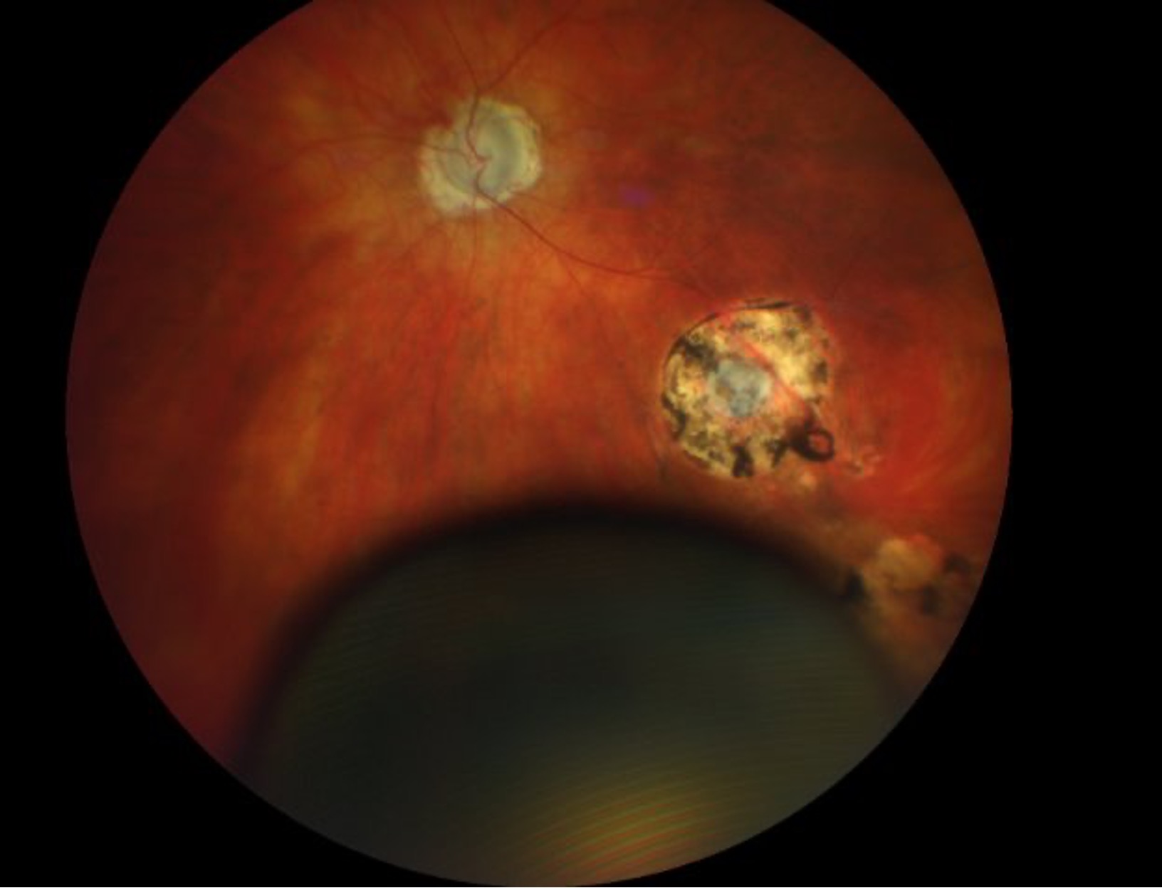
Posterior segment imaging is important to rule out differential diagnosis (ex. endophthalmitis)(6) and detect co-morbid posterior segment pathology prior to cataract extraction.

Treatment/Management/Guidelines

Early presentation is a crucial prognostic factor; one study of 50 patients with lens-induced glaucoma found that vision was preserved if medical and surgical management occurred within 2 weeks of symptom onset(7). Definitive treatment is cataract extraction(2). Concurrent trabeculectomy can be considered if the patient presents seven or more days after symptom onset, especially in settings where postoperative medical IOP management may be unavailable. In our case, cataract extraction was not a treatment option due to pre-existing no light perception in the affected eye.

Medical management includes cycloplegics for pain management, aggressive topical medications to lower IOP, and topical steroids to decrease inflammation. Patients should undergo postoperative dilated fundus examination and visual field testing to establish a new baseline and detect posterior pathology that was previously obscured by the hypermature cataract.


EPIDEMIOLOGY OR ETIOLOGY

  • Rare cause of secondary open-angle glaucoma
  • Cataract hypermaturity secondary to:
    • Trauma
    • Uncontrolled diabetes
    • Lack of access to healthcare and/or low vision potential that prevent earlier cataract extraction

SIGNS

  • Hypermature cataract
  • Elevated IOP (although can be low due to ciliary body shut own)
  • Tenderness
  • Open angle
  • Anterior chamber cell and flare
  • Corneal edema
  • Lens dislocation

SYMPTOMS

  • Unilateral Pain
  • Decreased Visual Acuity
  • Photophobia
  • Redness/Injection

TREATMENT/MANAGEMENT

  • CEIOL
  • Aggressive IOP Management
    • Topical ɑ2 agonist or ꞵ-Blocker
    • Systemic carbonic anhydrase inhibitors
  • Topical Steroids (prednisolone acetate 1%)

RELATED LINKS:


References
  1. Epstein DL, Jedziniak JA, Grant WM. Obstruction of aqueous outflow by lens particles and by heavy-molecular-weight soluble lens proteins. Invest Ophthalmol Vis Sci 1978;17(3):272-277. [PMID 627464]
  2. J MS, Thomas M, E FW, A OT. Phacolytic Glaucoma due to Morgagnian Cataract. Eyerounds.org 2018, https://EyeRounds.org/cases/258-phacolytic-glaucoma.htm
  3. Flocks M, Littwin CS, Zimmerman LE. Phacolytic glaucoma; a clinicopathologic study of one hundred thirty-eight cases of glaucoma associated with hypermature cataract. AMA Arch Ophthalmol 1955;54(1):37-45. [PMID 14375459]
  4. Gervasio K, Peck T. The Wills Eye Manual: Office and Emergency Room Diagnosis and Treatment of Eye Disease: Wolters Kluwer Health, 2021.
  5. Camacho M, Sayegh Y, Dubovy SR. Phacolytic Glaucoma Diagnosed by Cytopathology: A Clinicopathologic Case Series. Ocul Immunol Inflamm 2024;32(9):2100-2104. [PMID 38592742]
  6. Dhingra D, Grover S, Kapatia G, Pandav SS, Kaushik S. Phacolytic glaucoma: A nearly forgotten entity. Eur J Ophthalmol 2020;30(5):Np32-np35. [PMID 30950286]
  7. Sharanabasamma M, Vaibhav K. Management and Visual Outcome in Patients of Lens-induced Glaucomas at a Tertiary Eye Care Hospital in South India. J Curr Glaucoma Pract 2016;10(2):68-75. [PMID 27536050]

Suggested citation format

Al-Kaylani HM, Lovett Jr. EA, Oladele-Ajose A, Boese E. Phacolytic glaucoma in an eye with no light perception. EyeRounds.org February 24, 2026. Available from https://EyeRounds.org/cases/375-phacolytic-glaucoma.htm


Image Permissions:
Creative Commons

Ophthalmic Atlas Images by EyeRounds.org, The University of Iowa are licensed under a Creative Commons Attribution-NonCommercial-NoDerivs 3.0 Unported License.