Chief Complaint: 54-year-old female patient presents requesting new glasses.
History of Present Illness: Patient reports mild blurriness in both eyes (OU) at distance and near for several months. Rarely she experiences episodes of monocular diplopia in each eye when exposed to bright light.
Past Ocular History: She is unaware of any eye problems and has had no professional eye care in many years.
Medical History: Hypertension, migraine headaches and depression
Medications: None
Social and Family History: Noncontributory
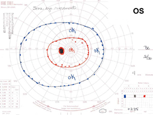
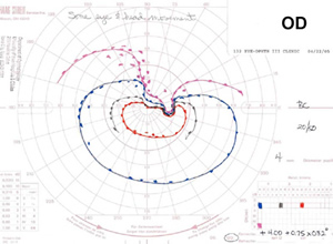
Course: Given the vague description of "blurred vision" and the visual field defect discovered on confrontation testing, further history was sought. The patient denied any symptoms of giant cell arteritis. She denied hypotension or acute onset of a visual field defect that might support suspicions of anterior ischemic optic neuropathy. Formal kinetic perimetry (Goldmann perimetry) was obtained prior to dilation (see Figure 1).
A: Goldmann visual field of the left eye (OS) reveals no abnormalities. |
B: Goldmann visual field of the right eye (OD) confirms a dense superior scotoma. |
 |
 |
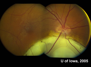
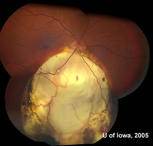
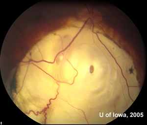
After visual field testing, the patient was dilated. Images of the fundus are shown below (see Figures 2 and 3).
| A: Fundus photograph, OD. Large inferonasal retinochoroidal coloboma. | B: Fundus photograph, OS. Unremarkable. |
 |
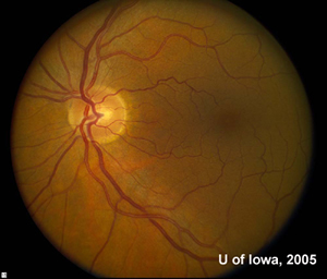 |
| A: Fundus photograph, OD. The extent of the coloboma is appreciated. | B: Fundus photograph, OD. Magnified view of retinochoroidal coloboma with associated staphyloma. |
 |
 |
Discussion: The patient was a poor historian and had no recollection of her ocular issues. We subsequently located medical records of this patient from 1955 which described the coloboma. Records from the early 1960’s reported reduced visual acuity is secondary to amblyopia.
Coloboma is usually caused by failure of the embryonic fissure to close in the 5th week of development. The coloboma can involve multiple layers of the globe, including the iris, lens, nerve, retina and retinal pigment epithelium (RPE). Failure of RPE development in the area of the coloboma may lead to hypoplasia of the choroid and sclera, resulting in staphyloma, as seen in this case.
Colobomas are usually inferonasal (typical coloboma), but may uncommonly occur in other locations (atypical coloboma). Colobomas may be unilateral or bilateral.
The CHARGE syndrome (Colobomas, Heart defects, coanal Atresia, Retardation of Growth and Ear abnormalities) may be associated with colobomas, especially bilateral colobomas. A variety of chromosomal abnormalities may be associated with colobomas. In terms of visual prognosis, foveal involvement is an important predictor of poor visual acuity. Even large colobomas which do not involve the fovea may exhibit relatively preserved visual acuity. Severe colobomas may be associated with microphthalmos.
Colobomas may be associated with a variety of ocular complications. Amblyopia and refractive error may be noted. Retinochoroidal colobomas may be associated with choroidal neovascularization and retinal detachment later in life.
EPIDEMIOLOGY
|
SIGNS
|
SYMPTOMS
|
TREATMENT
|
Tantri AP, Johnson AT: Retinochoroidal coloboma: 54-year-old female with blurred vision. Eyerounds.org. February 27, 2006; Available from: http://www.EyeRounds.org/cases/53-Retina-Choroidal-Coloboma-Visual-Field.htm.

Ophthalmic Atlas Images by EyeRounds.org, The University of Iowa are licensed under a Creative Commons Attribution-NonCommercial-NoDerivs 3.0 Unported License.