This site uses tracking information. Visit our privacy policy. Click to agree to this policy and not see this again.

Ophthalmology and Visual Sciences

Vogt-Koyanagi-Harada (VKH) Disease

Vogt-Koyanagi-Harada (VKH) Disease

Anthony P. Mai, BS; Charlene Tran, BS; Caroline W. Wilson, MD; Austin R. Fox, MD; H. Culver Boldt, MD
April 1, 2019

INITIAL PRESENTATION

Chief Complaint

Blurry vision and headaches

History of Present Illness

A 44-year-old Vietnamese woman presented to the emergency department with a 10-day history of progressive blurry vision in both eyes and a three-day history of severe headaches. Her central vision loss had not improved with a refraction by her optometrist. Her severe occipital headaches worsened with movement and were associated with generalized malaise, extreme fatigue, mild photophobia, and tearing. Acetaminophen partially alleviated the pain.

She had recently traveled to Vietnam but denied encountering sick contacts there. She denied jaw claudication, fevers, or weight changes. She denied skin rashes, hearing changes, tinnitus, dizziness, numbness, or tingling. She denied ever having tuberculosis. She had no history of prior vision problems, autoimmune conditions, or cancer.

Past Ocular History

  • History of cosmetic eyelid surgery (bilateral blepharoplasty) three years prior
  • No history of ocular trauma or disease

Past Medical History

None

Medications

Acetaminophen as needed

Allergies

No known drug allergies

Family History

No history of eye disease or autoimmune disease

Social History

She immigrated from Vietnam several years prior to presentation.  She is married and has three children. She works at a nail salon. She does not consume tobacco products, alcohol, or illicit substances. She travels to Vietnam every six to twelve months.

Review of Systems

Negative except for what is detailed in the history of present illness

OCULAR EXAMINATION

Visual Acuity with/without correction (Snellen)

  • Right eye (OD): 20/300 (no improvement with pinhole)
  • Left eye (OS): 20/60-2+2 (no improvement with pinhole)

Ocular Motility/Alignment

Full extraocular movements in both eyes (OU)

Intraocular Pressure (IOP): (Tonopen)

  • OD: 12 mmHg
  • OS: 14 mmHg

Pupils

  • OD: 4 mm in dark, 3 mm in light, no relative afferent pupillary defect (RAPD)
  • OS: 4 mm in dark, 3 mm in light, no RAPD

Confrontation visual fields: (Count fingers)

  • OD: Central scotoma
  • OS: Total inferotemporal defect

External

Normal on both sides

Slit lamp exam

  • Lids/lashes: Normal OU
  • Conjunctiva/sclera: Clear and quiet OU
  • Cornea: 1+ punctate epithelial erosions, no keratic precipitates OU
  • Anterior chamber: Trace cell and flare and deep OU
  • Iris: Normal architecture OU
  • Lens: Clear OU

Dilated fundus examination (DFE)

  • Vitreous: Trace anterior vitreous cells OU
  • Disc:
    • OD: Grade 3 disc edema, hyperemic
    • OS: Grade 2-3 disc edema, hyperemic
  • Cup-to-disc ratio: 0.0 OU
  • Macula:
    • OD: 3+ cystoid macular edema (CME) and subretinal fluid (SRF) extending from the disc to the temporal macula. No lipid or exudates. Boggy-appearing choroid.
    • OS: 2+ CME and SRF extending from the disc through the fovea. 1-2+ linear lipid extending from disc towards fovea. Boggy-appearing choroid.
  • Vessels:
    • OD: Sheathing temporally
    • OS: Normal
  • Periphery:
    • OD: Cystic retinal tuft anterior to the equator at 10:30
    • OS: Shallow SRF anterior to the equator at 4:00

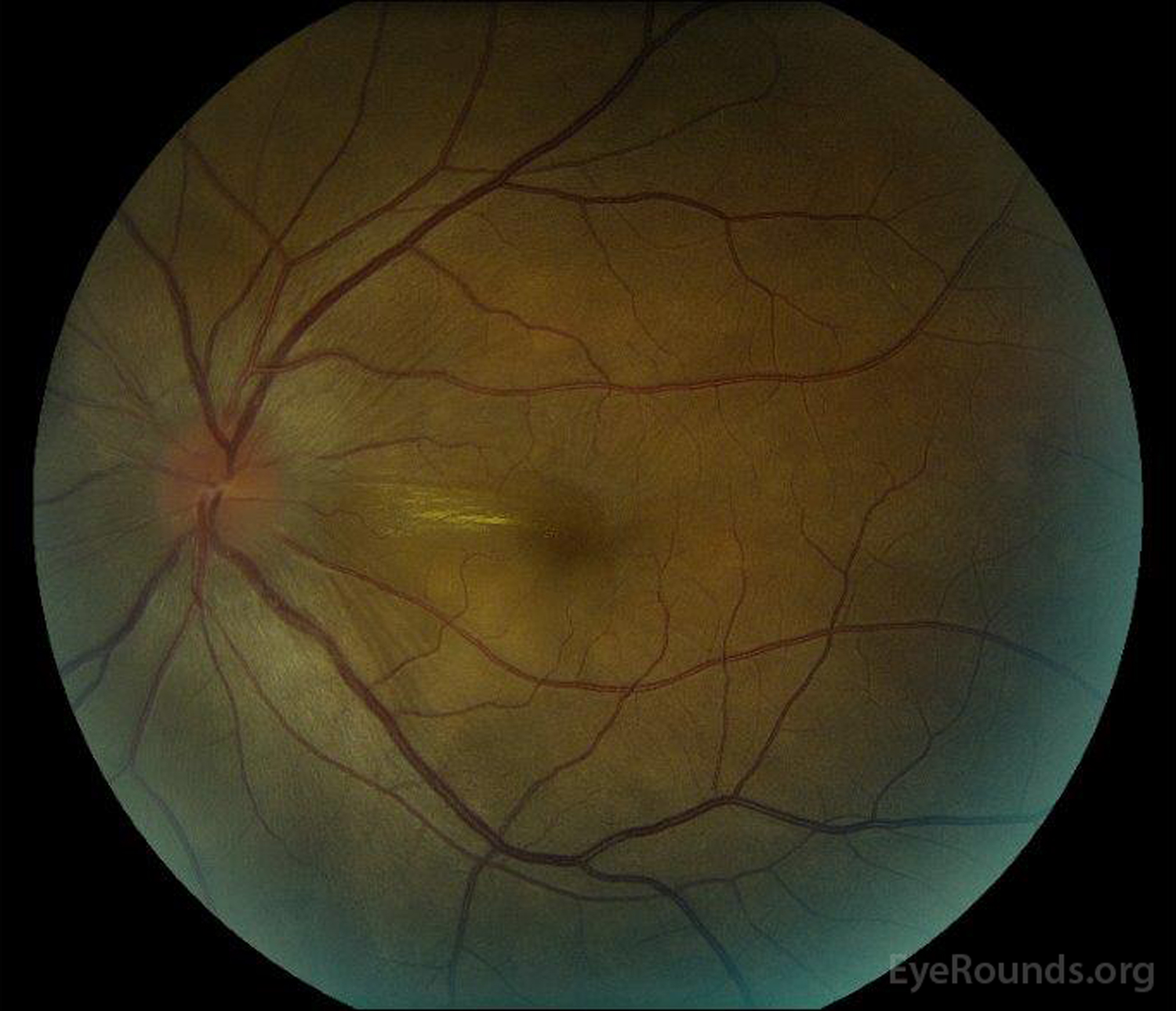
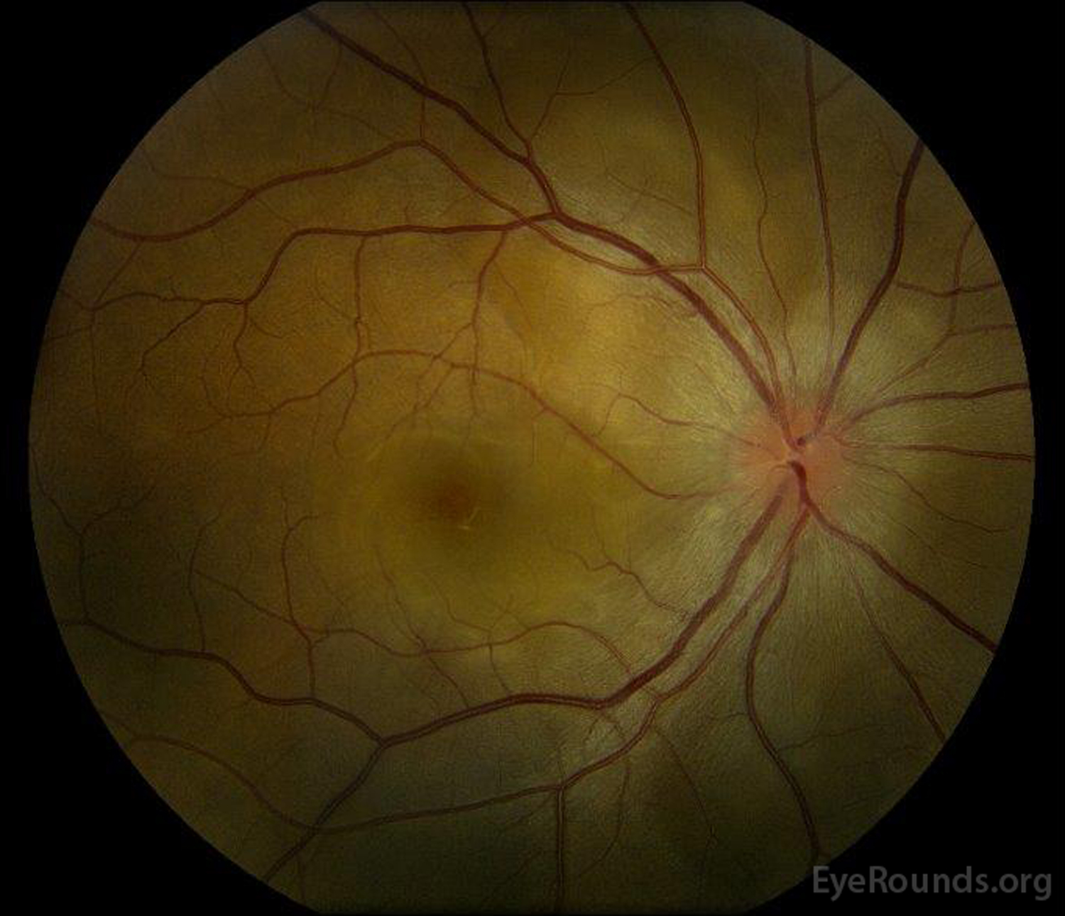
Figure 1: Color fundus photographs at presentation: (Left image) The right eye has disc edema and mild hyperemia as well as subretinal fluid extending from the disc temporally through the macula. There is also a focal serous retinal detachment superotemporal to the disc, along the superior arcade. (Right image) The left eye has disc edema and mild hyperemia, along with subretinal fluid extending from the disc to the macula and linear lipid deposits in the nasal macula. 

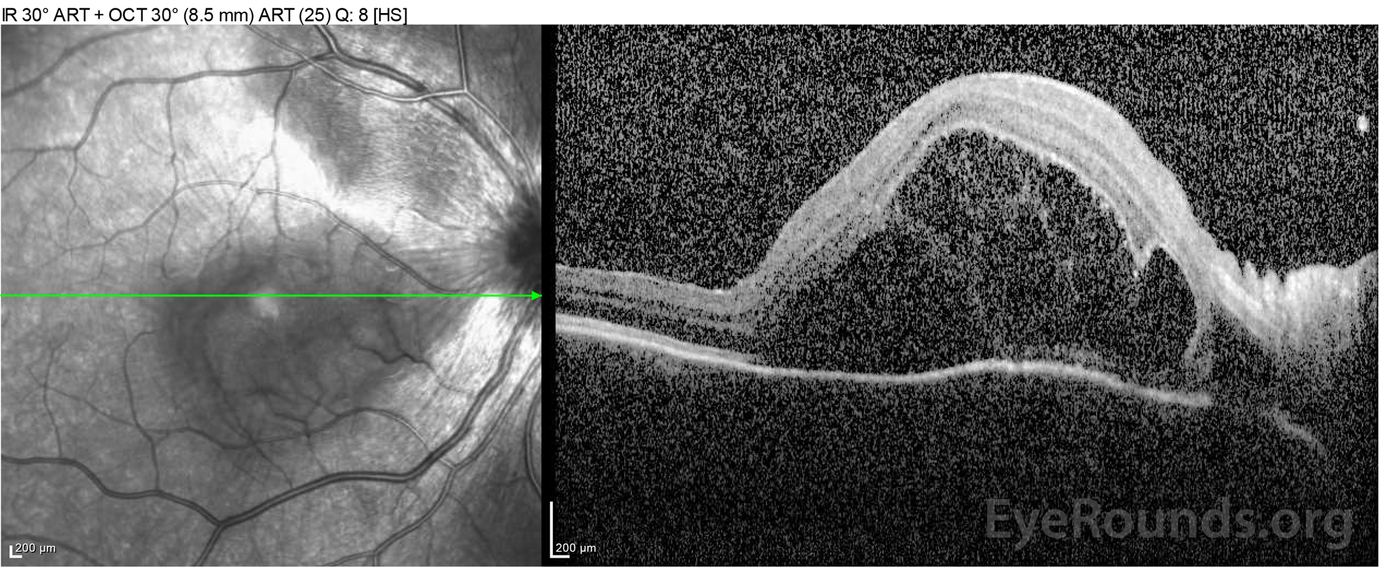
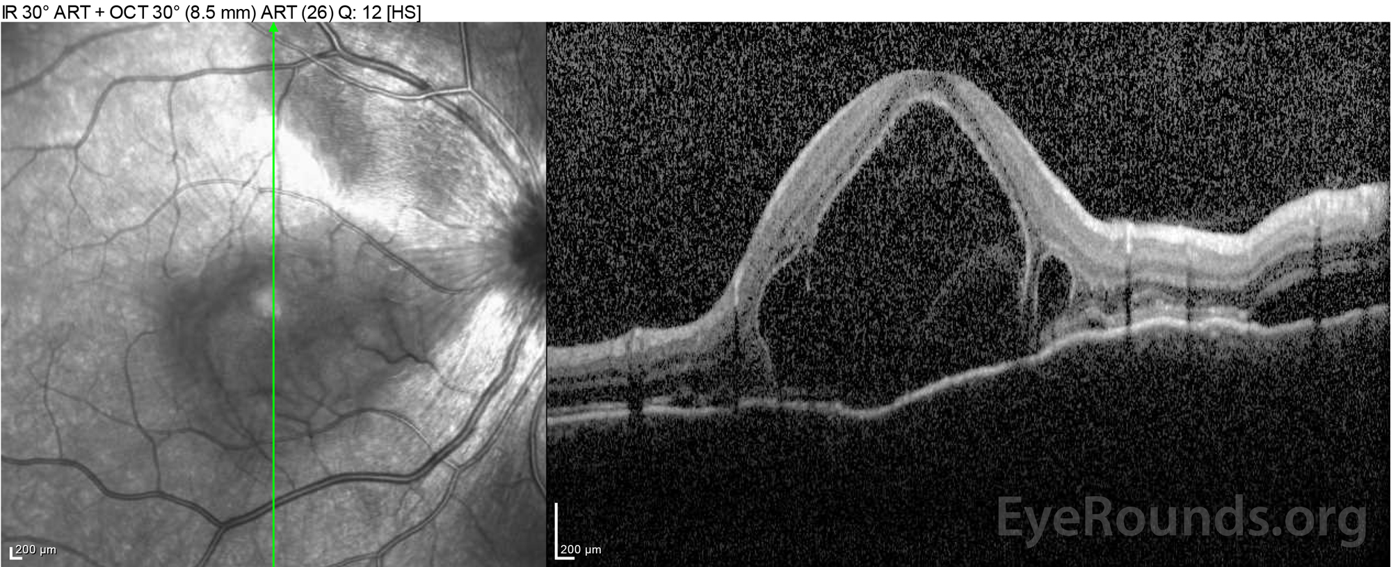
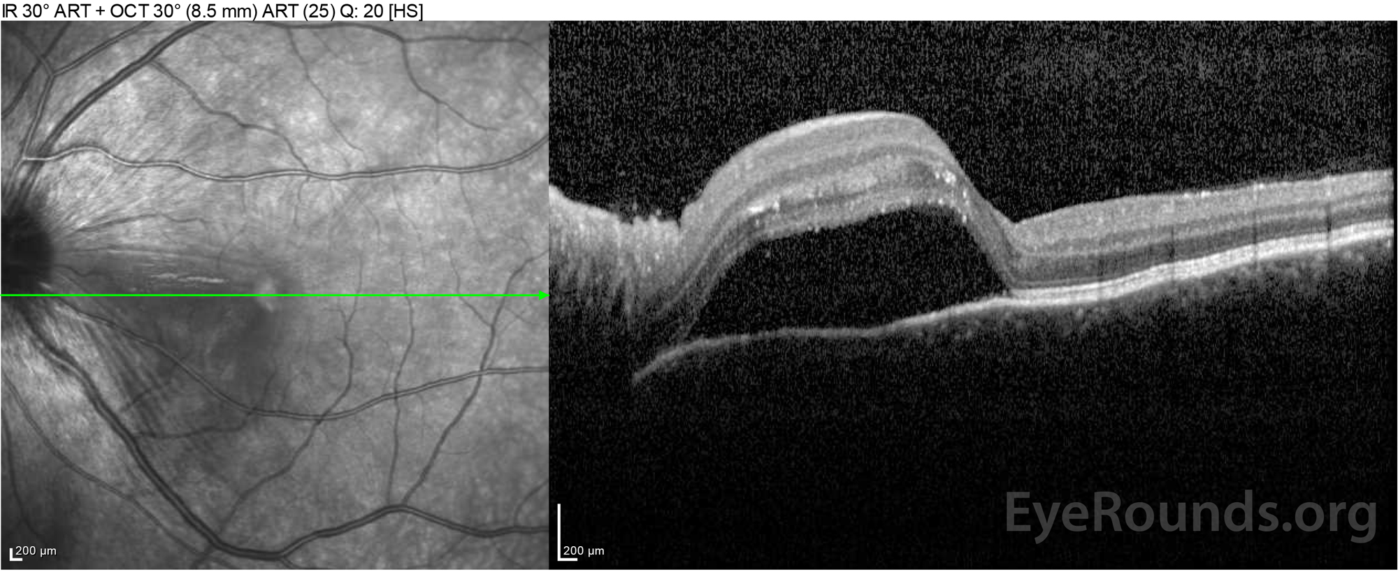
Figure 2: Optical coherence tomography (OCT) of the right eye (upper panels) shows a serous retinal detachment involving the fovea with extensive overlying intraretinal fluid, disruption of the outer retinal layers, and undulations of the thickened choroid. OCT of the left eye (bottom panels) shows a serous retinal detachment in the nasal macula extending up to the fovea.

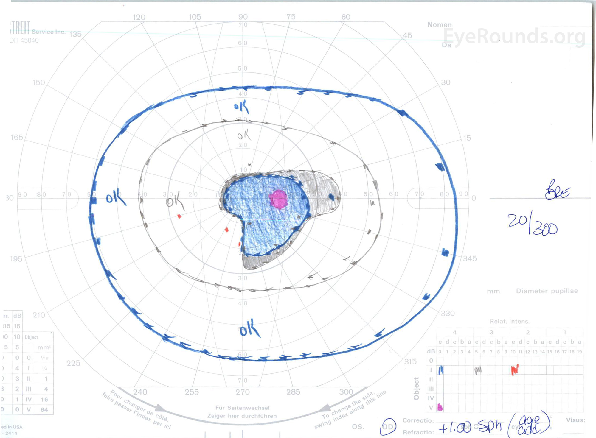
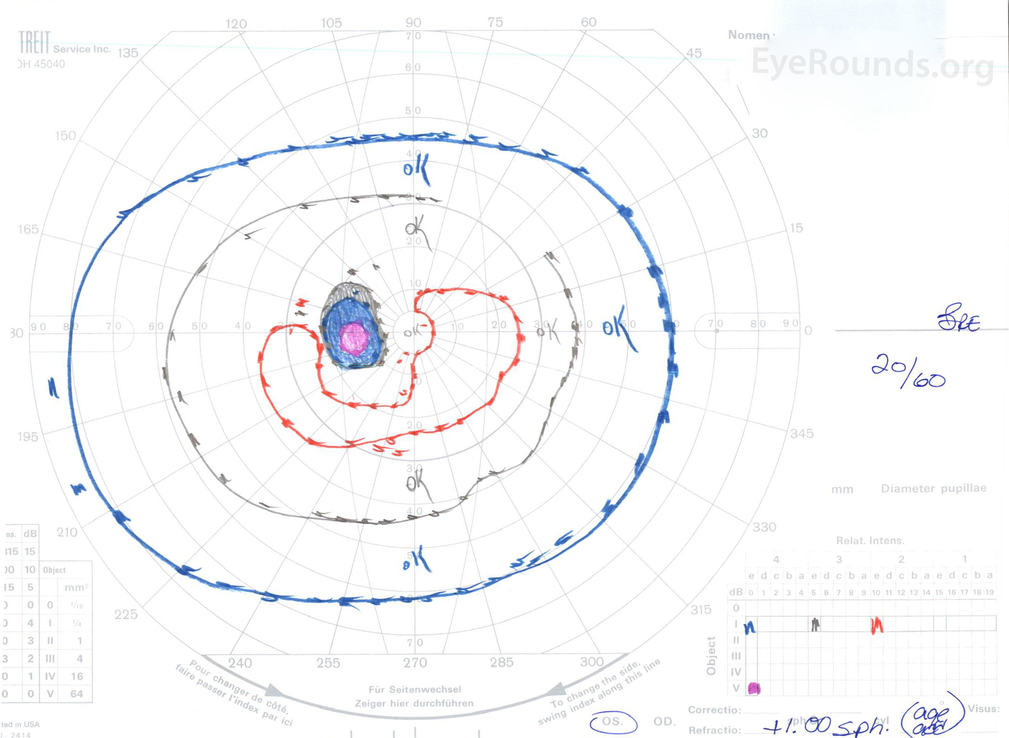
Figure 3: Goldman visual fields (GVF), OU. (Left image) OS shows an enlarged physiologic blind spot and mild central scotoma. (Right image) OD shows a moderately dense central scotoma incorporating the physiologic blind spot and extending inferotemporally.

B-scan: No signs of scleritis, mild vitreal opacities/cells inferiorly

Differential Diagnosis

WORK-UP

Complete blood count

White blood cell count: 4.9 K/mm3 (Ref: 3.7-10.5)
Red blood cell count 3.99 M/mm3 (Ref: 4.0-5.2)
Hemoglobin 11.6 g/dL (Ref: 11.9-15.5)
Hematocrit 35 % (Ref: 35-47)

Basic metabolic panel

Sodium 138 mEq/L (Ref: 135-145)
Potassium 4.3 mEq/L (Ref: 3.5-5.0)
Chloride 107 mEq/L (Ref: 95-107)
CO2 20 mEq/L (Ref: 22-29)
Blood urea nitrogen 16 mEq/dL (Ref: 10-20)
Creatinine 0.7 mg/dL (Ref: 0.5-1.0)

C-reactive Protein (CRP): <0.5 mg/dL  (Ref: <=0.5)
Erythrocyte sedimentation rate (ESR): 12 mm/Hr (Ref: 0-20)
Angiotensin--converting enzyme (ACE): 13 U/L (Ref: 8-52)
QuantiFERON-TB Gold: negative
Iron, blood 54 microgram/dL (Ref: 37-145)
Total iron binding capacity 379 microgram/dL (Ref: 250-425)

CLINICAL COURSE

The patient was initially evaluated by the emergency department given her complaints of new-onset severe headaches and vision loss. Brain computed tomography (CT) and magnetic resonance imaging (MRI) scans were unremarkable. ESR and CRP were within normal levels. The ophthalmology clinic evaluated her the following day and found bilateral serous retinal detachments and panuveitis. ACE and QuantiFERON-TB Gold labs were both negative.  She was diagnosed with Vogt-Koyanagi-Harada disease based on her clinical presentation and Asian descent. She was treated with 80mg of prednisone daily, acetaminophen as needed for headaches, and Vitamin D and calcium supplementation. Her headaches rapidly resolved, and her visual acuity steadily improved over the following two weeks. Her prednisone dosage was then tapered down to 40 mg over three weeks with continued resolution of symptoms and improvement in visual acuity.  She had no recurrence of headaches or worsening vision during the prednisone taper.  At her most recent appointment, she had tapered down to 5mg every other day, without return of symptoms.  Her visual acuity at that follow-up visit was 20/15-2 OD and 20/20+2 OS, and macular OCT showed full resolution of disc edema and serous retinal detachments in both eyes (Figure 4).

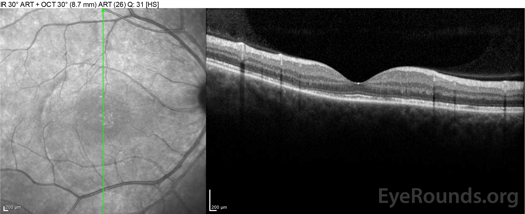
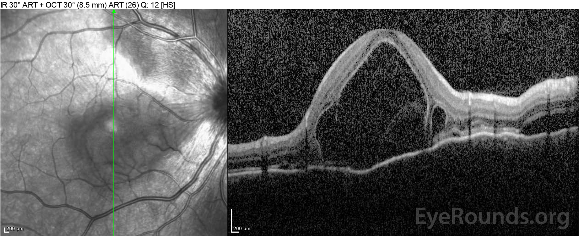
Figure 4: Optical coherence tomography showing subretinal fluid at baseline (top) and the course of resolution at one week (middle) and five weeks (bottom) while on a high-dose oral prednisone taper. Note the smoothing out of the choroidal undulations with treatment.

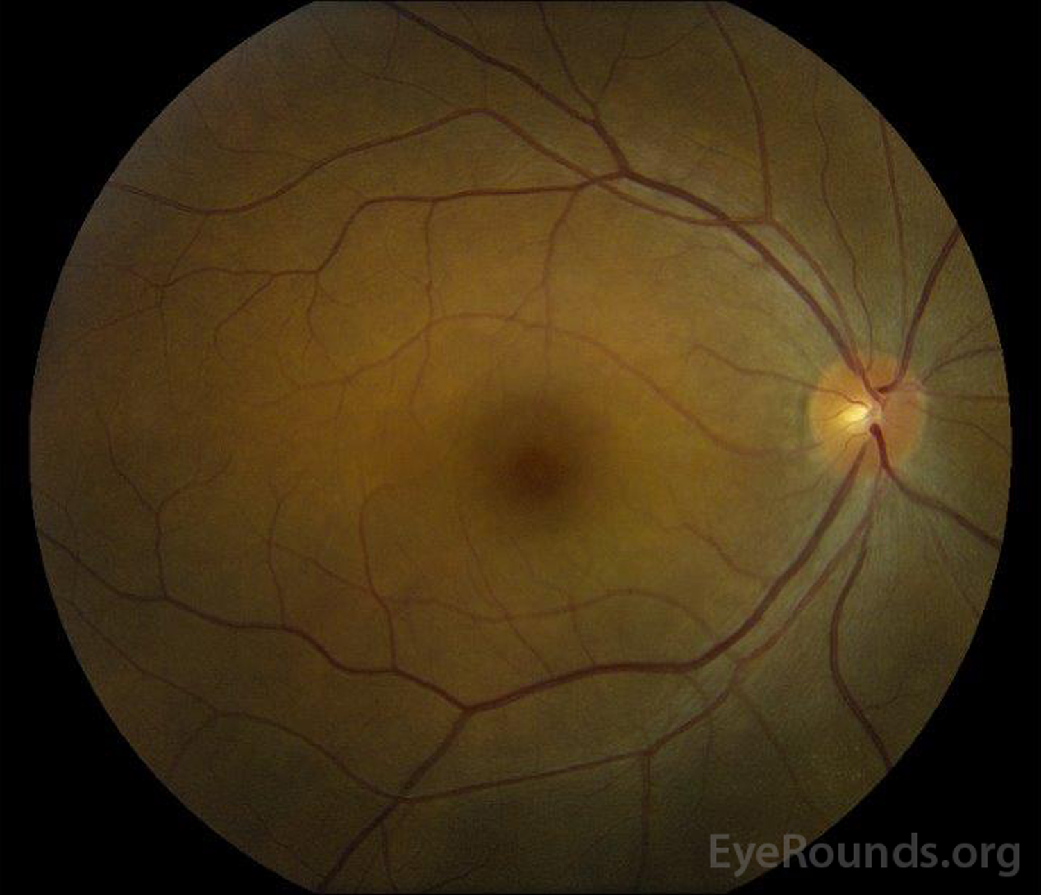
Figure 5: Color fundus photography of the right (A) and left (B) eyes during the convalescence phase demonstrating improvement in the subretinal fluid and disc edema.

DIAGNOSIS

Incomplete Vogt-Koyanagi-Harada Disease

DISCUSSION

Vogt-Koyanagi-Harada (VKH) Disease is a systemic autoimmune condition characterized by bilateral non-necrotizing granulomatous panuveitis associated with extraocular integumentary changes, such as poliosis and vitiligo, and inflammation affecting the uvea, inner ear, hair, and meninges. Harada disease is the isolated uveitis without the systemic signs or symptoms of VKH.

Etiology

The etiology of VKH disease is still largely unknown despite current research efforts. It is thought to be an acquired autoimmune disease involving T-cell-mediated hypersensitivity to melanocytic self-antigens, with an underlying genetic predisposition and possible microbial trigger [1]. Tyrosinase and tyrosinase-related peptides are melanocyte antigens that have been suggested as targets of autoimmune processes in VKH [2]. However, increased risk of VKH disease was not associated with the tyrosinase gene family, according to one study [3].

Because of the increased prevalence among certain ethnic and gender groups, there is thought to be a genetic predisposition in the pathogenesis of VKH. Multiple genes, including human leukocyte antigen (HLA) and interleukin (IL) genes, have been associated with VKH in different ethnic populations [4]. HLA receptors are major histocompatibility complexes in humans that present peptides to the immune system. HLA-DR1, HLA-DR4, HLA-DRB1*0405, and HLA-DRw53 are several haplotypes found in patients with VKH disease [5,6]. HLA-DR4 is more common in Japanese and Hispanic people, while HLA-DRB1*0405 is more frequent in Korean and Middle Eastern patients [7]. Both the HLA-DR4 and HLA-DRB1*0405 alleles are found in Vietnamese patients [8]. The HLA-DRB1 receptor binds to melanocyte antigens in varying capacities. Despite these associations, genetic testing is not recommended at this time.

Given the usual prodromal symptoms that accompany VKH, including fever, headaches, meningismus and tinnitus, an inciting viral etiology has been suggested as a trigger for VKH onset through mechanisms of molecular mimicry in genetically predisposed patients. Cytomegalovirus envelope glycoprotein H has significant amino acid homology to the tyrosinase peptide, and CMV infection may trigger VKH through molecular mimicry (i.e., recognition by HLA-class II receptors) [9]. Ebstein-bar virus (EBV) has also been implicated. However, there has been no definitive evidence regarding a viral etiology of VKH, and it remains unclear what triggers the VKH autoimmune response [1].

Pathophysiology

There are four classic phases of VKH that can have variable presentations: prodromal, acute uveitic, convalescent, and chronic-recurrent. Histopathologic changes typically begin in the acute phase [7].

The acute uveitic phase is characterized by bilateral uveal thickening secondary to granulomatous inflammation. The granulomas consist of lymphocytes, macrophages, and granule-filled epithelioid and giant cells [10,11]. Although the epithelioid cells were previously believed to be altered melanocytes, a follow-up immunohistochemical study suggested an origin from tissue macrophages instead [1,12]. Granulomas filled with epithelioid histiocytes, termed Dalen-Fuchs nodules, can often be seen between the retinal pigment epithelium (RPE) and Bruch's membrane. The uveal granulomatous inflammation leads to choroidal thickening and exudative retinal detachments filled with proteinaceous fluid. In addition, pleocytosis (i.e., increased cell count) may be present in the anterior chamber and vitreous [7,11].

The convalescent phase is identified by depigmentation of the choroid and extraocular areas, including the periocular skin and hair.  A depigmented choroid set against a pale optic nerve gives the impression of a "sunset-glow" fundus, which is a classic feature of this phase of VKH [10].  In addition, Dalen-Fuchs nodules become more prominent underneath the RPE in the convalescent phase [6].

The chronic-recurrent phase is characterized by decreased choroidal thickness, resolution of serous retinal detachments, chronic mild vitritis, and recurrent granulomatous anterior segment inflammation. Choroidal neovascularization (CNV) and subretinal fibrosis may develop during this phase and are indicators of severe disease progression [7,11]. Cataracts and secondary glaucoma are other complications of long-standing or recurrent inflammation in this phase [10].

Epidemiology

VKH is prevalent in races with darker skin pigment, especially Asians, South Americans, Middle Easterners, and Native Americans. VKH disease accounts for >10% of uveitis in these populations [5-7,10,11,13,14].  Only 1-4% of uveitis cases are thought to be secondary to VKH disease in the United States (7). In the United States, most cases of VKH have been found to affect individuals of Asian, Hispanic, and/or Native American decent . Interestingly, VKH disease rarely affects Africans despite their dark pigmentation [15]. The incidence of VKH disease varies greatly between racial subgroups in neighboring countries [1,7,10] .  For instance, Korea's incidence of VKH is only 2%, much lower than that found in Japan and China [16].

 VKH has a typical onset of 20 to 50 years of age [17-20]; however, studies suggest that 3.1-13.4% of VKH cases are pediatric patients and 10% of cases are ≥65-years-old [1]. Classically, VKH is thought to have a predilection for the female gender, and while most studies show that VKH disproportionately affects women, a few studies have shown a male predisposition or no gender predisposition [21].

Signs/Symptoms

As aforementioned, the four stages of VKH disease are prodromal, uveitic, convalescent, and chronic recurrent. Each stage exhibits distinct clinical features.

  • Prodromal: This initial stage may present as a influenza -like illness with predominantly constitutional symptoms, such as headache, dizziness, fever, fatigue, and/or nausea. Neurological symptoms of meningitis, cranial nerve palsies, and optic neuritis, as well as auditory symptoms of tinnitus, dysacusis, and vertigo have been reported [5-7, 10].  Photophobia, blurred vision, floaters, and/or eye pain usually begin within 48 hours of prodromal symptoms [11]. The prodromal phase typically lasts from a few days to weeks.
  • Acute Uveitic: This stage includes blurry vision, photophobia, conjunctival injection, and eye pain. There may be mild anterior uveitis that at first appears non-granulomatous.  Unilateral onset typically transitions to bilateral involvement within 1-2 weeks. Granulomatous anterior uveitis with mutton-fat keratic precipitates may develop. Posterior exam findings may include optic nerve edema and hyperemia [22], multifocal areas of choroiditis, multiple areas of serous retinal detachments localized to the posterior fundus, choroidal thickening, radiating chorioretinal folds, and vitritis [6,7,10] . Serous retinal detachments may form a cloverleaf pattern in the posterior fundus and could progress to extensive bullous detachments in severe cases [11]. Acute inflammatory glaucoma has been associated with this phase of the disease and can present with a shallow anterior chamber secondary to ciliary body edema, mimicking acute-angle closure [5]. The duration of the acute uveitic phase depends on the prompt diagnosis and management.
  • Chronic Uveitic or Convalescent: This stage typically develops several weeks after the acute phase and is characterized by vitiligo (e.g., face, hands, shoulders, or back), poliosis, and alopecia [7]. Depigmentation near the corneal limbus, known as Sugiura's sign, can be seen one month after disease onset [11]; however, this sign is rarely seen outside of the Japanese population [1]. Choroidal depigmentation usually occurs over a few months and results in the bright orange-red color of the choroid and the classic "sunset glow fundus."  Sunset glow fundus is thought to be the most important and predictive in the diagnosis of chronic VKH [23]. Well-defined, round, nummular chorioretinal scars can form in the mid-periphery. The chronic uveitic phase typically lasts several months.
  • Chronic-recurrent: This stage is characterized by recurrent episodes of granulomatous anterior uveitis with mutton fat keratic precipitates, iris nodules, iris depigmentation, posterior synechiae, posterior subcapsular cataracts, secondary glaucoma, choroidal neovascular membranes, and, ultimately, subretinal fibrosis and nummular chorioretinal atrophy [23,24]. The chronic phase typically develops at least six months after initial presentation. The serous retinal detachments present during the acute and convalescent phases typically do not recur with aggressive corticosteroid treatment [11]. 

Diagnostic Criteria

The most recent diagnostic criteria, named the Revised Diagnostic Criteria (RDC) for VKH, were defined in 1999 at the First International Workshop on VKH [15]These are outlined in Table 1.  The RDC are useful in that they divide VKH into three different diagnostic categories based upon the disease phase during which a patient presents: complete, incomplete, and probable.  This categorization of disease allows for appropriate and early management in "probable" disease that may help prevent progression to "complete" disease.

Work-up for other causes of ocular inflammation, both infectious and auto-inflammatory, are essential.  These can include erythrocyte sedimentation rate (ESR), C-reactive protein (CRP), quantiferon-Gold testing for tuberculosis, rapid plasma reagin (RPR) for syphilis, angiotensin-converting enzyme (ACE) and a chest x-ray for sarcoidosis, antinuclear antibody (ANA), and p-/c-ANCA. Also, a history of recent ocular trauma or intraocular surgery must be noted and likely suggests sympathetic ophthalmia (SO) as the more likely diagnosis given the very similar presentation and pathophysiology shared between SO and VKH [25]. 

To support a diagnosis of VKH in equivocal cases, a lumbar puncture can be performed to look for lymphocytic and monocytic pleocytosis; however, this is rarely employed clinically. Eighty percent of patients have pleocytosis in the cerebrospinal fluid (CSF) within one week and 97% have pleocytosis within three weeks. Increased levels of immune cells can last up to eight weeks after disease onset [1,11] . The T-cell surface marker profiles are similar between the CSF and the aqueous humor but different from the blood. This suggests the ability of CSF to accurately reflect uveal inflammation in VKH disease [26].

Table 1. Revised Diagnostic Criteria for Vogt-Koyanagi-Harada Disease

*From Table 1 in (15).

"Complete Vogt-Koyanagi-Harada disease (criteria 1 to 5 must be present)

  1. No history of penetrating ocular trauma or surgery preceding the initial onset of uveitis.
  2. No clinical or laboratory evidence suggestive of other ocular disease entities.
  3. Bilateral ocular involvement (a or b must be met, depending on the stage of disease when the patient is examined).
    1. Early manifestations of disease.
      1. There must be evidence of a diffuse choroiditis (with or without anterior uveitis, vitreous inflammatory reaction, or optic disk hyperemia), which may manifest as one of the following:
        1. Focal areas of subretinal fluid, or
        2. Bullous serous retinal detachments.
      1. With equivocal fundus findings; both of the following must be present as well:
        1. Focal areas of delay in choroidal perfusion, multifocal areas of pinpoint leakage, large placoid areas of hyperfluorescence, pooling within subretinal fluid, and optic nerve staining (listed in order of sequential appearance) by fluorescein angiography, and
        2. Diffuse choroidal thickening, without evidence of posterior scleritis by ultrasonography.
    1. Late manifestations of disease.
      1. History suggestive of prior presence of findings from 3a, and either both (2) and (3) below or multiple signs from (3):
      2. Ocular depigmentation (either of the following manifestations is sufficient): (a) Sunset glow fundus, or (b) Sugiura sign.
      3. Other ocular signs:
        1. Nummular chorioretinal depigmented scars, or
        2. Retinal pigment epithelium clumping and/or migration, or
        3. Recurrent or chronic anterior uveitis.
  4. Neurological/auditory findings (may have resolved by time of examination).
    1. Meningismus (malaise, fever, headache, nausea, abdominal pain, stiffness of the neck and back, or a combination of these factors; headache alone is not sufficient to meet definition of meningismus, however), or
    2. Tinnitus, or
    3. Cerebrospinal fluid pleocytosis.
  5. Integumentary finding (not preceding onset of central nervous system or ocular disease).
    1. Alopecia, or
    2. Poliosis, or
    3. Vitiligo.

Incomplete Vogt-Koyanagi-Harada disease (criteria 1 to 3 and either 4 or 5 must be present)

  1. No history of penetrating ocular trauma or surgery preceding the initial onset of uveitis, and
  2. No clinical or laboratory evidence suggestive of other ocular disease entities, and
  3. Bilateral ocular involvement.
  4. Neurologic/auditory findings; as defined for complete Vogt-Koyanagi-Harada disease above, or
  5. Integumentary findings; as defined for complete Vogt-Koyanagi-Harada disease above.

Probable Vogt-Koyanagi-Harada disease (isolated ocular disease; criteria 1 to 3 must be present)

  1. No history of penetrating ocular trauma or surgery preceding the initial onset of uveitis.
  2. No clinical or laboratory evidence suggestive of other ocular disease entities.
  3. Bilateral ocular involvement as defined for complete Vogt-Koyanagi-Harada disease above. "

Testing/Laboratory Work-up

In the initial workup of VKH, one should consider obtaining the following tests:

  • Optical coherence tomography (OCT): In the acute uveitic phase, OCT will likely show significant choroidal thickening and serous retinal detachments. The subretinal fluid accumulations may have septations believed to be fibrin membranes and inflammatory products, creating a lobular structure that can also be seen on fluorescein angiography.  In the convalescent phase, OCT can detect areas of retinal thinning following resolved inflammation after corticosteroid treatment [1, 6, 7, 11].
  • B-scan ultrasonography: In the acute phase, ultrasonography may show diffuse posterior choroidal thickening, posterior scleral thickening, retinal detachments, and vitreous opacities [1, 7, 11]. Ciliary effusions may be observed with ultrasound biomicroscopy [5]. This test is also useful for ruling out posterior scleritis.
  • Fluorescein angiography (FA): Classically, FA reveals multifocal choroidal hypofluorescent dots at the early phase followed by multiple focal hyperfluorescent areas with diffuse leakage in the late phase [6, 7].  The dye leaks through the RPE and accumulates in the subretinal space surrounding the hyperfluorescent dots. FA can be diagnostically useful when VKH disease presents without extraocular symptoms. Optic disc hyperfluorescence and window defects caused by atrophic chorioretinal scars may be seen in the mid-periphery [1, 5, 11]. FA in the chronic-recurrent stage of VKH disease shows nonspecific window defects due to RPE damage, choroidal neovascularization, and subretinal fibrosis [1, 11].
  • Indocyanine green (ICG) angiography: Early phase ICG depicts hyperfluorescent stromal vessels that indicate choroidal vasculopathy and hypofluorescent dark dots that correspond to granulomas and delayed patchy filling of choroidal vasculature [27]. The late phase reveals fuzzy stromal vascular patterns and diffuse choroidal hyperfluorescence. Disc hyperfluorescence is suggestive of severe disease. ICGA can detect subclinical choroidal inflammation in very early stages or even after systemic therapy [1, 5-7].
  • Lumbar puncture: Pleocytosis in the cerebrospinal fluid is present in the majority of VKH patients. Lumbar puncture should be performed early in the disease course since pleocytosis may resolve

Treatment/Management/Guidelines

Treatment goals in VKH include early diagnosis and suppression of active inflammation, along with prevention of recurrent inflammation and vision-threatening complications, such as glaucoma, bullous retinal detachment, and choroidal neovascularization.   

Systemic corticosteroid treatment is the preferred therapy for VKH disease, especially during the acute uveitic stage. It has been shown that the route of corticosteroid administration (oral versus intravenous) does not impact the visual acuity or occurrence of visually-significant complications in the treatment of acute VKH [29].  For severe disease, the suggested protocol is intravenous methylprednisolone administration for three days followed by oral high-dose prednisone treatment. In mild-moderate disease, high-dose oral prednisone may be sufficient at 1-2mg/kg/day. The steroid dose should be slowly tapered over approximately six months to prevent recurrence [30]. Aggressive early treatment, alongside serial FA testing showing disappearance of dye leakage through the RPE, may help to prevent further disease progression, recurrence, and extraocular manifestations [7]. Topical steroids and cycloplegics can decrease cells in the anterior chamber and vitreous humor.

Intravitreal and sub-Tenon injections of triamcinolone have been used for short-term control of intraocular inflammation during the acute or recurrent phases; these local therapies should be considered in the case of recalcitrant disease and in patients who poorly tolerate the unfavorable systemic side effects of steroids given the extended steroid taper.  Intravitreal anti-VEGF injections are sometimes used for control of choroidal neovascularization and in cases of persistent foveal serous retinal detachments [1, 5, 7, 25].

Steroid-sparing agents including antimetabolites, calcineurin inhibitors, biologics, TNF-alpha inhibitors, or cytotoxic agents can be used to treat VKH and should be carefully monitored, often in coordination with a rheumatology service [1]. There has been ongoing discussion regarding the use of non-steroidal immunosuppressive agents as first-line therapy for VKH disease.  However, a recent study revealed no differences in outcomes between early first-line immunomodulatory treatment (IMT) and prednisone treatment alone [28]. Further, immunosuppressive and biologic therapies are expensive and require careful pre-treatment evaluation as well as frequent follow-up with blood work to assess for serious side effects.

In the chronic-recurrent stage, frequent recurrence may suggest resistance to corticosteroid therapy and suggests need for steroid-sparing immunomodulatory treatment [1, 6, 11].  The preferred agent for steroid-resistant recurrence or steroid-intolerance is cyclosporine [7]. Infliximab, rituximab, adalimumab, and interferon alpha-2a are biologic agents that have also been used to treat refractory uveitis in VKH disease.

To treat the anterior uveitis often associated with acute VKH, topical steroids (e.g., prednisolone acetate 1%) and topical cyloplegia (e.g., cyclopentolate 1% or atropine 1%) should be prescribed depending on the degree of anterior chamber inflammation.

Ocular complications are commonly associated with VKH disease. Given the multiple stages and variety of presentations in which a patient can present with VKH, treatment may be delayed in many cases. In severe forms of VKH and in recurrences, intraocular inflammation may be difficult to control and may result in structural damage. Over 50% of patients develop related complications, including cataract, secondary glaucoma, choroidal neovascular membranes, subretinal fibrosis, or a combination of these (Figure 6) [31].

Venn diagram showing complications in VKH patients. (Used with permission from Am J Ophthalmol. 2001;131(5):599-606

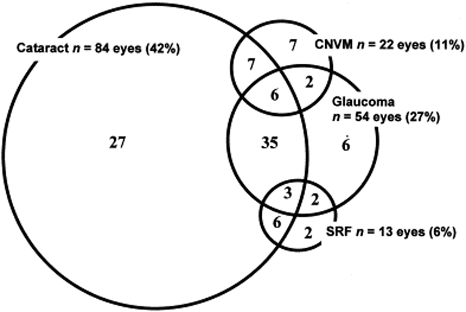
Figure 6: Venn diagram showing complications in VKH patients. (Used with permission from Am J Ophthalmol. 2001;131(5):599-606 [31])

EPIDEMIOLOGY AND ETIOLOGY
  1. Typical age of onset: 20 to 50 years
  2. Female > male
  3. Predisposition in populations with pigmented skin, particularly Asian, Middle Eastern, Hispanic, and Native Americans
  4. Genetic risks: HLA-DR1, HLA-DR4, HLA-DRB1*0405 alleles
  5. Possible autoimmune targeting of melanocyte antigens
  6. Possible viral molecular mimicry as inciting factor in autoinflammatory activation
SIGNS
  1. Prodromal: Pleocytosis in CSF, meningitis, vertigo
  2. Uveitic: Bilateral granulomatous anterior or multifocal uveitis, multifocal serous retinal detachments in both eyes, optic nerve head edema and hyperemia
  3. Convalescent: Alopecia, poliosis, sunset glow fundus, Sugiura sign (limbal depigmentation), Dalen-Fuchs nodules below the RPE
  4. Chronic recurrent: Recurrence of anterior uveitis, subretinal fibrosis, choroidal neovascular membranes, multifocal chorioretinal scars, secondary glaucoma, cataracts
SYMPTOMS
  1. Influenza-like prodrome with headache, fever, malaise, nausea, and/or vomiting
  2. Hearing loss, tinnitus and dysacusis
  3. Neck stiffness
  4. Rapidly worsening blurry vision involving both eyes
  5. Eye redness, pain, floaters, and/or photophobia
TREATMENT/MANAGEMENT
  1. High-dose systemic corticosteroids (intravenous vs oral) for the acute phase with slow taper
  2. Immunosuppressive agents (cyclosporine) and biologics (infliximab) for recurrent disease refractory to corticosteroids
  3. Topical steroids (prednisolone acetate 1%) and cycloplegics (cyclopentolate 1% or atropine 1%) for anterior uveitis and ciliary spasm
  4. Intravitreal triamcinolone and/or intravitreal anti-VEGF injections for control of inflammation, choroidal neovascular membrane, and/or persistent subfoveal fluid
References
  1. Du L, Kijlstra A, Yang P. Vogt-Koyanagi-Harada disease: Novel insights into pathophysiology, diagnosis and treatment. Prog Retin Eye Res 2016;52:84-111.  https://PubMed.gov/26875727. DOI: 10.1016/j.preteyeres.2016.02.002
  2. Yamaki K, Gocho K, Hayakawa K, Kondo I, Sakuragi S. Tyrosinase family proteins are antigens specific to Vogt-Koyanagi-Harada disease. J Immunol 2000;165(12):7323-7329.  https://PubMed.gov/11120868
  3. Horie Y, Takemoto Y, Miyazaki A, Namba K, Kase S, Yoshida K, Ota M, Hasumi Y, Inoko H, Mizuki N, Ohno S. Tyrosinase gene family and Vogt-Koyanagi-Harada disease in Japanese patients. Mol Vis 2006;12:1601-1605.  https://PubMed.gov/17200659
  4. Ng JY, Luk FO, Lai TY, Pang CP. Influence of molecular genetics in Vogt-Koyanagi-Harada disease. J Ophthalmic Inflamm Infect 2014;4:20.  https://PubMed.gov/25097674. DOI: 10.1186/s12348-014-0020-1
  5. Bowling B. Uveitis. Kanski's Clinical Ophthalmology New York, New York: Elsevier; 2016; chapter 11; p. 395-465.
  6. Yeh PT YC, Yang CH, Lin CP. Nonrhegmatogenous Retinal Detachment. In: Schachat AP SS, Hinton DR, Wilkinson CP, Wiedemann P,, editor. Ryan's Retina. New York: Elsevier; 2018; chapter 99; p. 1828-1849.
  7. Goto H RK, Rao N. Vogt–Koyanagi–Harada Disease. In: Schachat AP SS, Hinton DR, Wilkinson CP, Wiedemann P, editor. Ryan's Retina. New York, New York: Elsevier; 2018; chapter 78; p. 1505-1515.
  8. Riddington L, Hall AJ, Tait B, Nicholson I, Varney M. Vogt-Koyanagi-Harada syndrome in patients of Vietnamese ancestry. Aust N Z J Ophthalmol 1996;24(2):147-149.  https://PubMed.gov/9199747
  9. Sugita S, Takase H, Kawaguchi T, Taguchi C, Mochizuki M. Cross-reaction between tyrosinase peptides and cytomegalovirus antigen by T cells from patients with Vogt-Koyanagi-Harada disease. Int Ophthalmol 2007;27(2-3):87-95.  https://PubMed.gov/17253112. DOI: 10.1007/s10792-006-9020-y
  10. Freund BK SD, Mieler WF, Yannuzzi LA. Inflammation. The Retinal Atlas. New York,  New York: Elsevier 2017; chapter 4; p. 279-398.
  11. Rao N. Vogt-Koyanagi-Harada Disease. In: J YMaD, editor. Ophthalmology. New York, New York: Elsevier; 2014; chapter 7.17; p. 761-763.
  12. Rao NA, Xu S, Font RL. Sympathetic ophthalmia. An immunohistochemical study of epithelioid and giant cells. Ophthalmology 1985;92(12):1660-1662.  https://PubMed.gov/4088616
  13. Nussenblatt RB. Vogt-Koyanagi-Harada Syndrome. In: Whitcup RBNaSM, editor. Uveitis: Fundamentals and Clinical Practice. 4th Edition ed: Elsevier; 2010; chapter Chapter 24.
  14. Read RW, Holland GN, Rao NA, Tabbara KF, Ohno S, Arellanes-Garcia L, Pivetti-Pezzi P, Tessler HH, Usui M. Revised diagnostic criteria for Vogt-Koyanagi-Harada disease: report of an international committee on nomenclature. Am J Ophthalmol 2001;131(5):647-652.  https://PubMed.gov/11336942
  15. Chung H, Choi DG. Clinical analysis of uveitis. Korean J Ophthalmol 1989;3(1):33-37.  https://PubMed.gov/2795939. DOI: 10.3341/kjo.1989.3.1.33
  16. Abu El-Asrar AM, Al-Kharashi AS, Aldibhi H, Al-Fraykh H, Kangave D. Vogt-Koyanagi-Harada disease in children. Eye (Lond) 2008;22(9):1124-1131.  https://PubMed.gov/17479116. DOI: 10.1038/sj.eye.6702859
  17. Martin TD, Rathinam SR, Cunningham ET. Prevalence, clinical characteristics, and causes of vision loss in children with Vogt-Koyanagi-Harada disease in South India. Retina 2010;30(7):1113-1121.  https://PubMed.gov/20168275. DOI: 10.1097/IAE.0b013e3181c96a87
  18. Forster DJ, Green RL, Rao NA. Unilateral manifestation of the Vogt-Koyanagi-Harada syndrome in a 7-year-old child. Am J Ophthalmol 1991;111(3):380-382.  https://PubMed.gov/2000916
  19. Yamamoto Y, Fukushima A, Nishino K, Koura Y, Komatsu T, Ueno H. Vogt-koyanagi-harada disease with onset in elderly patients aged 68 to 89 years. Jpn J Ophthalmol 2007;51(1):60-63.  https://PubMed.gov/17295144. DOI: 10.1007/s10384-006-0379-0
  20. Wang Y, Chan CC. Gender differences in vogt-koyanagi-harada disease and sympathetic ophthalmia. J Ophthalmol 2014;2014:157803.  https://PubMed.gov/24734166. DOI: 10.1155/2014/157803
  21. Nakao K, Abematsu N, Mizushima Y, Sakamoto T. Optic disc swelling in Vogt-Koyanagi-Harada disease. Invest Ophthalmol Vis Sci 2012;53(4):1917-1922.  https://PubMed.gov/22408010. DOI: 10.1167/iovs.11-8984
  22. Rao NA, Gupta A, Dustin L, Chee SP, Okada AA, Khairallah M, Bodaghi B, Lehoang P, Accorinti M, Mochizuki M, Prabriputaloong T, Read RW. Frequency of distinguishing clinical features in Vogt-Koyanagi-Harada disease. Ophthalmology 2010;117(3):591-599, 599.e591.  https://PubMed.gov/20036008. DOI: 10.1016/j.ophtha.2009.08.030
  23. Veerappan M, Fleischman D, Ulrich JN, Stinnett SS, Jaffe GJ, Allingham RR. The Relationship of Vogt-Koyanagi-Harada Syndrome to Ocular Hypertension and Glaucoma. Ocul Immunol Inflamm 2017;25(6):748-752.  https://PubMed.gov/27438521. DOI: 10.1080/09273948.2016.1189578
  24. Baltmr A, Lightman S, Tomkins-Netzer O. Vogt-Koyanagi-Harada syndrome - current perspectives. Clin Ophthalmol 2016;10:2345-2361.  https://PubMed.gov/27932857. DOI: 10.2147/OPTH.S94866
  25. Kitaichi N, Matoba H, Ohno S. The positive role of lumbar puncture in the diagnosis of Vogt-Koyanagi-Harada disease: lymphocyte subsets in the aqueous humor and cerebrospinal fluid. Int Ophthalmol 2007;27(2-3):97-103.  https://PubMed.gov/17211585. DOI: 10.1007/s10792-006-9016-7
  26. Oshima Y, Harino S, Hara Y, Tano Y. Indocyanine green angiographic findings in Vogt-Koyanagi-Harada disease. Am J Ophthalmol 1996;122(1):58-66.  https://PubMed.gov/8659599
  27. Read RW, Yu F, Accorinti M, Bodaghi B, Chee SP, Fardeau C, Goto H, Holland GN, Kawashima H, Kojima E, Lehoang P, Lemaitre C, Okada AA, Pivetti-Pezzi P, Secchi A, See RF, Tabbara KF, Usui M, Rao NA. Evaluation of the effect on outcomes of the route of administration of corticosteroids in acute Vogt-Koyanagi-Harada disease. Am J Ophthalmol 2006;142(1):119-124.  https://PubMed.gov/16815259. DOI: 10.1016/j.ajo.2006.02.049
  28. Rubsamen PE, Gass JD. Vogt-Koyanagi-Harada syndrome. Clinical course, therapy, and long-term visual outcome. Arch Ophthalmol 1991;109(5):682-687.  https://PubMed.gov/2025171
  29. Urzua CA, Velasquez V, Sabat P, Berger O, Ramirez S, Goecke A, Vásquez DH, Gatica H, Guerrero J. Earlier immunomodulatory treatment is associated with better visual outcomes in a subset of patients with Vogt-Koyanagi-Harada disease. Acta Ophthalmol 2015;93(6):e475-480.  https://PubMed.gov/25565265. DOI: 10.1111/aos.12648
  30. Read RW, Rechodouni A, Butani N, Johnston R, LaBree LD, Smith RE, Rao NA. Complications and prognostic factors in Vogt-Koyanagi-Harada disease. Am J Ophthalmol 2001;131(5):599-606.  https://PubMed.gov/11336934
Suggested citation format:

Mai AP, Tran C, Wilson CW, Fox AR, Boldt HC. Vogt-Koyanagi-Harada (VKH) Disease. EyeRounds.org. April 1, 2019. Available from https://eyerounds.org/cases/284-vogt-koyanagi-harada.htm


last updated: 04/1/2019

Image Permissions:
Creative Commons

Ophthalmic Atlas Images by EyeRounds.org, The University of Iowa are licensed under a Creative Commons Attribution-NonCommercial-NoDerivs 3.0 Unported License.