This site uses tracking information. Visit our privacy policy. Click to agree to this policy and not see this again.

Ophthalmology and Visual Sciences

Diagnosis and Management of Combined Hamartoma of the Retina and Retinal Pigment Epithelium (CHRRPE) in Pediatric Patients

A three-part case series of pediatric patients with CHRRPE and discussion of the association with neurofibromatosis type 2

Diagnosis and Management of Combined Hamartoma of the Retina and Retinal Pigment Epithelium (CHRRPE) in Pediatric Patients

A three-part case series of pediatric patients with CHRRPE and discussion of the association with neurofibromatosis type 2
Ella Gehrke, BS, ScM; Margaret Strampe, MD; Elaine Binkley, MD; Lindsay De Andrade, MD
August 3, 2023

Case #1

INITIAL PRESENTATION

Chief Complaint: Newly identified retinal lesion following eye exam

History of Present Illness: Patient is a 3-year-old male, initially presenting as a referral after an eye exam. The patient’s father provided the history and denied new symptoms of vision change, drifting, or crossing of the eyes. He did report noticing that the patient needed to increase the brightness level when looking at phone screens. The patient received his first pair of glasses for amblyopia two weeks prior to presentation. Since that time, the patient’s father reported good compliance with glasses. The patient has no known symptoms of hearing loss or other medical problems.

Past Ocular History: Amblyopia; treated with prescription lenses, not worn at time of visit but parents report good compliance.

Past Medical History:

  • Born at term via spontaneous vaginal birth
  • Meeting developmental milestones

Medications: None

Allergies: No known allergies

Family History:

  • Older sister patches for strabismus
  • No known genetic conditions


OCULAR EXAMINATION

  • Visual Acuity without correction
    • Distance: Right eye (OD) 20/20, Left Eye (OS) 20/40
    • Near: OD 20/20, OS 20/80
  • Intraocular Pressure (iCare)
    • OD: 12 mm Hg
    • OS: 15 mm Hg
  • Pupil Exam No relative afferent pupillary defect (RAPD) in Both Eyes (OU)
  • Ocular Motility/Alignment: No nystagmus or strabismus
  • External Exam: Normal OU
  • Slit Lamp Exam
    • Lids/lashes: Normal OU
    • Conjunctiva/sclera: Clear and quiet OU
    • Cornea: Clear OU
    • Anterior chamber: Deep and quiet OU
    • Iris: Normal architecture
    • Lens: Normal OU
  • Dilated fundus examination (DFE)
    • Disc: Normal OU
    • Macula:
      • Normal OD
      • Grey/brown pigmented lesion in central macula with foveal involvement OS
    • Vessels: Normal OU
    • Periphery: Normal OU
  • Testing
  • MRI of the brain without contrast. Prior to presentation, MRI revealed globe flattening in an axial view (left panel, red arrows) and a partially empty sella in saggital view (right panel, red arrow).
    Figure 1: Fundus photo of the left eye demonstrated a grey/brown pigmented lesion in the central macula with foveal involvement. The vessels, optic disc, and visible mid-periphery were grossly normal.
    Color fundus photography, right (A) and left (B) eyes. Both eyes demonstrated grade IV optic disc edema with cotton wool spots, peripapillary hemorrhages, and retinal folds. In addition, scattered flame and dot-blot hemorrhages were seen in both eyes.
    Figure 2: : Optical coherence tomography (OCT) of the left eye showed disorganization and thickening of the retinal laminations centrally involving the fovea. There was a pre-retinal highly reflective membrane overlying the area of thickened retina. There was no intraretinal or subretinal fluid.

    Case #2

    INITIAL PRESENTATION

    Chief Complaint: Newly identified retinal lesion following routine eye exam

    History of Present Illness: Patient is a 4-year-old male. His initial presentation followed a failed vision screening, where the patient was instructed to start patching the left eye for possible amblyopia in the right eye. Patient’s parents report significant difficulty with patching, with behavior changes and falling asleep. At routine follow up with his local eye doctor, a retinal abnormality was noted and the patient was referred. The patient had no symptoms of hearing loss and parents did not have any specific vision concerns.

    Past Ocular History: Deprivation amblyopia, right eye, attempting patching

    Past Medical History:

    • Born at term via spontaneous vaginal birth
    • Meeting developmental milestones

    Medications: None

    Allergies: No known allergies

    Family History:

    • Brother patches for lazy eye, without strabismus
    • No known genetic conditions


    OCULAR EXAMINATION

    • Visual Acuity without correction
      • Distance: OD 20/60 +1, OS 20/20
      • Near: OD 20/60, OS 20/20
    • Intraocular Pressure (iCare)
      • OD: 16 mm Hg
      • OS: 16 mm Hg
    • Pupil Exam
      • OD: 0.3 log unit RAPD
      • OS: No RAPD
    • Ocular Motility/Alignment: No nystagmus or strabismus
    • External Exam: Normal OU
    • Slit Lamp Exam
      • Lids/lashes: Normal OU
      • Conjunctiva/sclera: Clear and quiet OU
      • Cornea: Clear OU
      • Anterior chamber: Deep and quiet OU
      • Iris: Normal architecture
      • Lens: Normal OU
    • Dilated fundus examination (DFE)
      • Disc: Central gliosis OD, normal OS
      • Macula: Large pigmented lesion OD, normal OS
      • Vessels: Gliosis over inferior arcade causing superior and temporal dragging of the vessels OD, normal OS
      • Periphery: Normal OU
    • Cycloplegic Refraction (Retinoscopy)
      Sphere Cylinder Axis
      Right +1.75 Sphere
      Left +1.00 +1.25 090
    • Testing
    • Color fundus photography, right (A) and left (B) eyes. Both eyes demonstrated grade IV optic disc edema with cotton wool spots, peripapillary hemorrhages, and retinal folds. In addition, scattered flame and dot-blot hemorrhages were seen in both eyes.
      Figure 3: Fundus photo of the right eye demonstrated a pigmented lesion in the macula. There was an epiretinal membrane (ERM) overlying the inferior arcade causing superior and temporal dragging of the vessels.
    • Optical Coherence Tomography (OCT)
    • Color fundus photography, right (A) and left (B) eyes. Both eyes demonstrated grade IV optic disc edema with cotton wool spots, peripapillary hemorrhages, and retinal folds. In addition, scattered flame and dot-blot hemorrhages were seen in both eyes.
      Figure 4: OCT of the right eye showed a central lesion with distortion and thickening of the inner and outer retina and an overlying epiretinal membrane. There was no intraretinal or subretinal fluid.

    Case #3

    INITIAL PRESENTATION

    Chief Complaint: Newly identified retinal lesion following development of blurry vision

    History of Present Illness: Patient is a 7-year-old male who was referred for newly noted retinal abnormalities identified after reporting blurry vision since start of the school year. He had also been experiencing headaches located at the back of his head that started after school, occurring several times per week. The headaches were relieved with sleep or rest and were not associated with nausea or vomiting. The patient had no history of hearing loss.

    Past Ocular History: Color blindness, congenital

    Past Medical History:

    • Term birth
    • Crohn’s disease

    Medications: Mesalamine (Crohn’s disease)

    Allergies: No known allergies

    Family History:

    • Crohn’s disease; mother and maternal grandmother
    • Color blindness; paternal grandfather


    OCULAR EXAMINATION

    • Visual Acuity without correction
      • Distance
        • OD: 20/70 -2 (pinhole 20/70, with correction 20/80-1)
        • OS: 20/40 (pinhole 20/20-3, with correction 20/20)
      • Near
        • OD: 20/30-1+1 (20/50+1 with correction)
        • OS: 20/20 (20/20 with correction)
    • Intraocular Pressure (iCare)
      • OD: 20 mm Hg
      • OS: 21 mm Hg
    • Pupil Exam
      • OU No RAPD
    • Color Exam Complete red green and strong Deutan colorblindness
    • Ocular Motility/Alignment: 7 prism diopters exophoria at near. Ortho at distance in all gazes
    • External Exam: Normal OU
    • Slit Lamp Exam
      • Lids/lashes: Normal OU
      • Conjunctiva/sclera: Clear and quiet OU
      • Cornea: Clear OU
      • Anterior chamber: Deep and quiet OU
      • Iris: Normal architecture
      • Lens: Normal OU
    • Dilated fundus examination (DFE)
      • Disc: Gliosis over optic nerve OD, normal OS
      • Macula: Large inferior pigmented lesion with dragging of vessels and overlying gliosis OD, normal OS
      • Vessels: Vessels dragged to macula OD, normal OS
      • Periphery: Normal OU
    • Cycloplegic Refraction (Retinoscopy)
      Sphere Cylinder Axis
      Right -0.50 +0.50 180
      Left -1.50 +.50 180
    • Testing
    • Color fundus photography, right (A) and left (B) eyes. Both eyes demonstrated grade IV optic disc edema with cotton wool spots, peripapillary hemorrhages, and retinal folds. In addition, scattered flame and dot-blot hemorrhages were seen in both eyes.
      Figure 5: Fundus photo of the right eye demonstrated a pigmented lesion in the macula associated with overlying gliotic membrane (ERM) and dragging of vessels.
      OCT of the right eye showed a central lesion with thickening and disorganization of the retinal layers and gliosis over the optic nerve.  There was no subretinal or intraretinal fluid..
      Figure 6: OCT of the right eye showed a central lesion with thickening and disorganization of the retinal layers and gliosis over the optic nerve. There was no subretinal or intraretinal fluid.

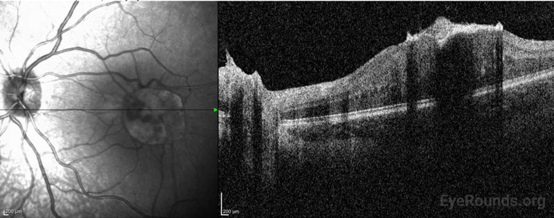
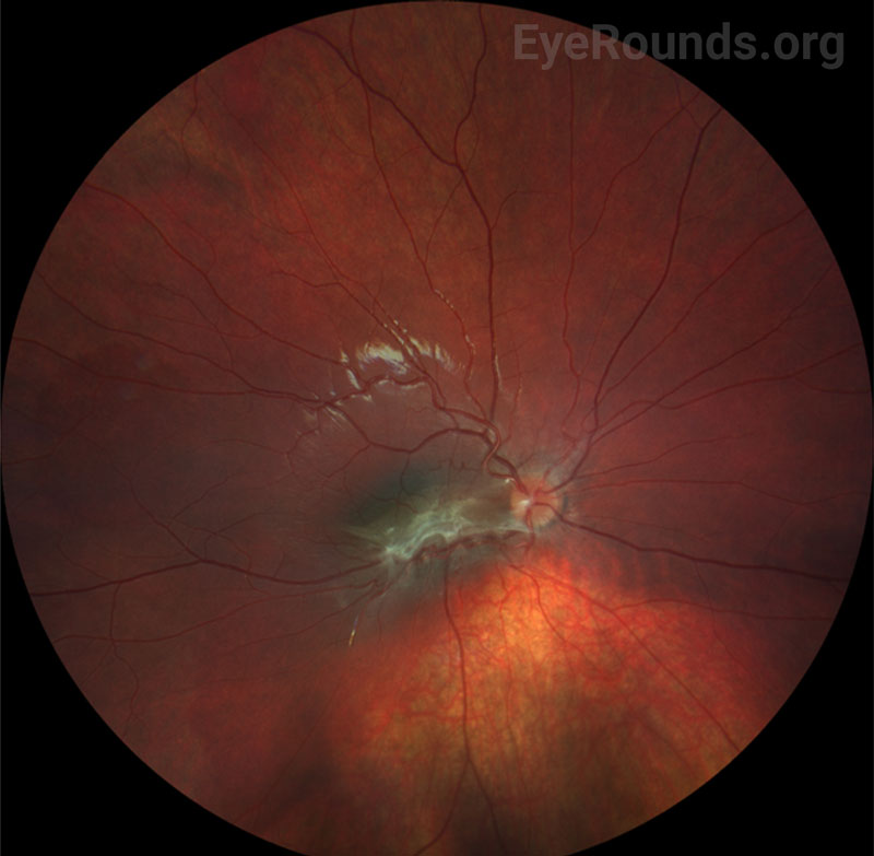
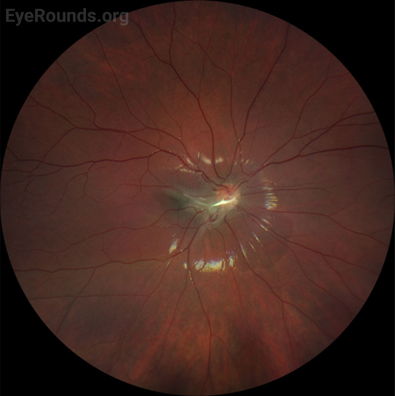
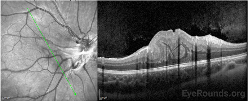
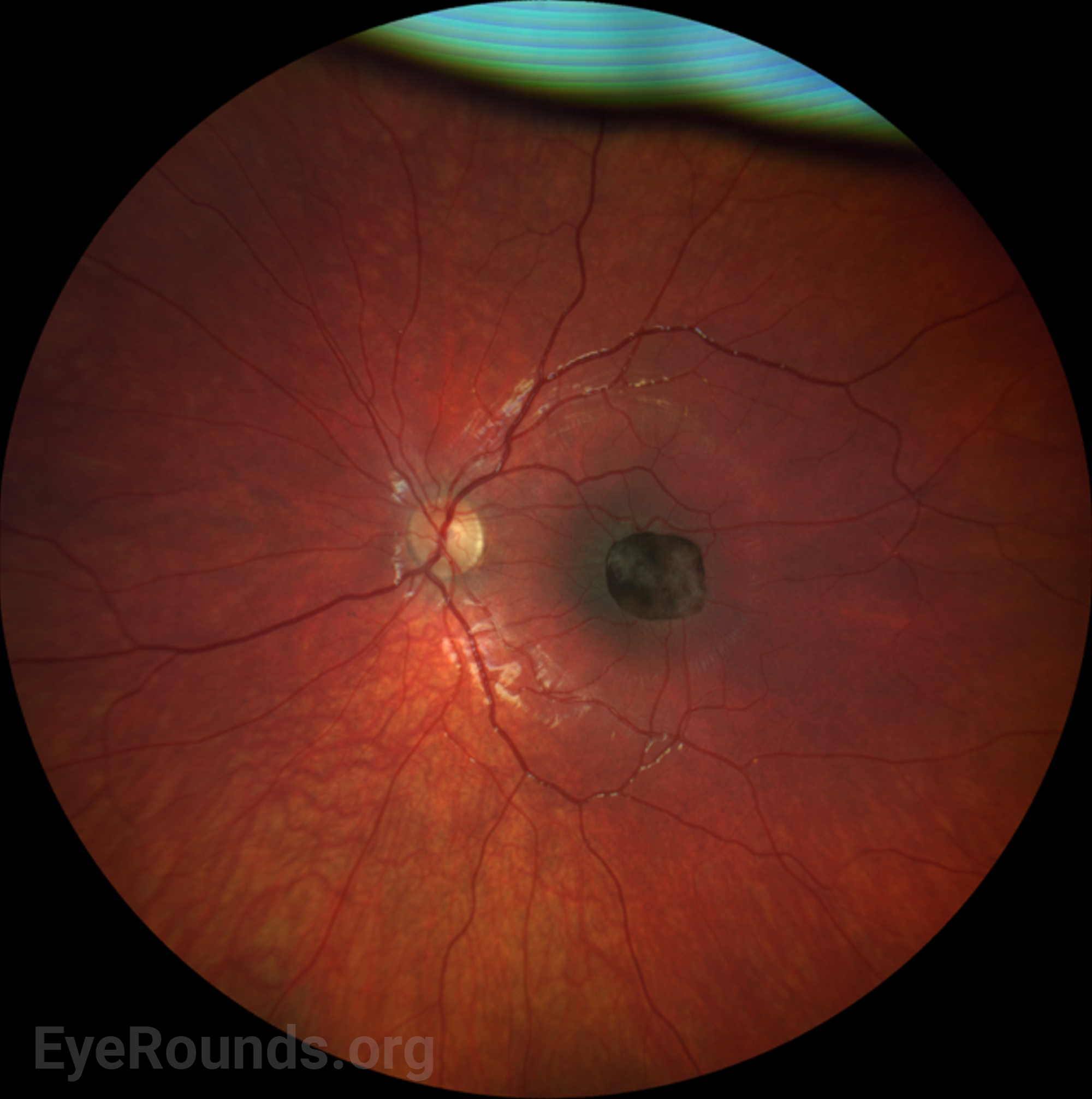
      DIAGNOSIS: Combined hamartoma of the retina and retinal pigmented epithelium (CHRRPE)


      CLINICAL COURSE

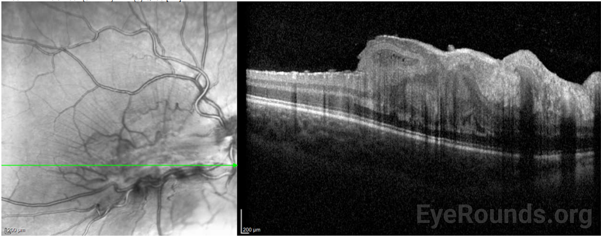
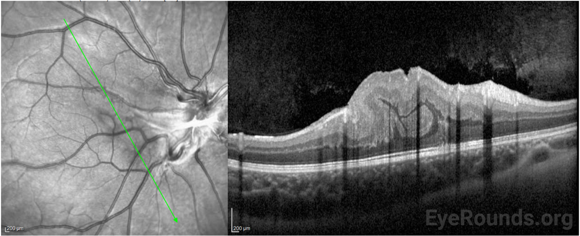
      A diagnosis of CHRRPE was made in each of the above cases as seen on fundus photography and confirmed using OCT. There was no known family history of neurofibromatosis or associated hearing loss in any of the patients, but due to the close association of CHHRPE with neurofibromatosis type 2 (NF2), it was recommended that all patients undergo formal genetic evaluation. Two of the three described patients followed with genetics for formal genetic testing. At that time, brain and spinal MRI (Magnetic Resonance Imaging) were ordered. Normal orbits and optic nerves were appreciated in both patients and no NF2-associated lesions were observed. Additionally, there were no variants in the NF2 gene of either patient. However, a NF2 diagnosis cannot be entirely ruled out due to the possibility of mosaicism, which has been demonstrated in NF2.[1]

      To date of publication, each of the described patients has remained stable. They have been evaluated every 6 months for management of amblyopia and routine counseling regarding monocular safety precautions. Patching for overlying deprivation amblyopia was attempted in each of the cases with varying degrees of success. Monocular safety precautions were strongly emphasized to each family to prevent injury to the unaffected eye.

      Pars plana vitrectomy with membrane peel was deferred in each of these cases given their stability but may be considered in the future if worsening epiretinal membrane causes a decline in acuity.


      DISCUSSION

      Combined hamartoma of the retina and retinal pigment epithelium (CHRRPE) is a rare benign proliferation of retinal glial cells and retinal pigment epithelium (RPE) as well as vascular tissue, that distorts the retina.[3] The mean age of diagnosis is 15 years old and les.[2] It is important to distinguish CHRRPE from malignant lesions such as retinoblastoma or melanoma. Unlike choroidal melanoma, CHRRPE is uniformly pigmented and possesses clearly demarcated borders.

      Although benign, the clinical manifestations of CHRRPE can include amblyopia, strabismus, vision loss, and metamorphopsia in patients.[3] Decreased vision and monocular strabismus are the most commonly reported symptoms, but as many as 2 out of 3 patients are asymptomatic at presentation.[4,5] Severity of vision loss is dependent on the location of the tumor. Macular involvement, as discussed in the associated cases, can significantly impact visual acuity (VA). CHRRPE with macular involvement have an average BCVA (best corrected visual acuity) of 20/320 compared to extramacular CHRRPE, which has an average BCVA of 20/80.

      The long-term complications of CHRRPE, although rare, can include retinal detachment, vitreous and retinal hemorrhage, choroidal neovascularization, and retinoschisis.3 Routine follow-up with an ophthalmologist, protective eyewear, and amblyopia interventions are crucial for maintaining vision in the pediatric patient.

      Neurofibromatosis Type 2 Association

      CHRRPE can occur spontaneously and are generally unilateral in these cases. However retinal and RPE (Retinal Pigment Epithelium) tumors are strongly associated with neurofibromatosis type 2 (NF2). Neurofibromatosis type 1 (NF1) and type 2 (NF2) are phakomatoses that are characterized by tumor formation due to mutations in tumor-suppressing genes. NF2 is caused by mutations on the NF2 gene, a known tumor suppressor located on 22q12.[2] Inheritance of NF2 is typically autosomal dominant; however, de novo mutations have been documented as well as somatic mosaicism.[7,8,2]

      NF2 demonstrates complete penetrance, but there is phenotypic variability. Generally, diagnosis of NF2 follows the identification of bilateral vestibular schwannomas and ocular and cutaneous tumors presenting in the 2nd and 3rd decade of life. However, pediatric patients have been identified with clinical features of NF2. In a retrospective study, the most common presenting symptom in children and young adolescents with NF2 was ophthalmic abnormalities rather than symptoms related to vestibular schwannoma as is seen in adults.[9] More specifically, studies have demonstrated retinal hamartomas in up to 22% of NF2 carriers.[7] Additional features of NF2 include meningiomas, ependymomas, and posterior subcapsular cataracts, emphasizing the importance of the ocular examination in pediatric and adult patients with NF2.

      Examination and Diagnostic Testing

      On dilated fundoscopic exam, CHRRPE are typically solitary and unilateral lesions that typically appear as elevated and uniformly pigmented lesions with fanlike projections. They can have different amounts of RPE, glial, and vascular components and are often usually covered by a grey-white semi-translucent membrane.1 Multimodal imaging including optical coherence tomography (OCT), standardized ocular ultrasound, and fluorescein angiography are useful in establishing a diagnosis. Additionally, serial fundus photography can help to follow the lesions over time. OCT is well tolerated by the pediatric population and can aid in diagnosis. Characteristic OCT findings in patients with CHRRPE include retinal striae, disorganization of the retina, and epiretinal membrane over the lesion surface.[10] A characteristic peak in the outer plexiform layer, referred to as a “saw tooth” pattern, is routinely described in the literature and can be observable on OCT.[11] Fundus fluorescein angiography can be used in diagnosis and to evaluate potential complications of CHRRPE such as choroidal neovascularization.12 Genetic evaluation and MRI should be considered at time of diagnosis due to the described NF2 association.

      Treatment & Management

      The primary goal of patching in pediatric patients with any unilateral visual threat is to prevent development of amblyopia. Amblyopia is a disruption in the eye-to-brain connection that develops when an eye is not being used due to disease or a history of trauma. It is also important for clinicians to emphasize the importance of protective eye wear, which serves as both a physical protection for the unaffected eye and a corrective lens if needed. Surgery to treat progressing ERM may be warranted in some cases. Early surgical intervention with pars plana vitrectomy with membrane peel has been shown to produce stable vision and reduce the amblyopic effect in a small study of children; however, reports of visual improvement with surgical intervention have been variable.[2] Routine follow up is necessary to evaluate for epiretinal membrane progression and neovascularization.


      Differential Diagnosis:

      ETIOLOGY

      • CHRRPE is a benign ocular proliferation of retinal glial cells, retinal pigment epithelium, as well as vascular tissue3
      • Can occur sporadically or be inherited in association with Neurofibromatosis type 2

      SIGNS

      • Variably decreased visual acuity
      • Pigmented lesion with fimbriated borders and overlying grey-white semi-translucent epiretinal membrane
      • Vessel/foveal dragging in some cases
      • Retinal thickening/disorganization

      SYMPTOMS

      • Decreased visual acuity
      • Strabismus
      • Amblyopia
      • Metamorphopsia

      MANAGEMENT

      • Amblyopia prevention
      • Protective eyewear
      • Genetic testing and MRI brain because of NF2 association
      • Surgical removal of the epiretinal membrane may be beneficial for some patients
References
  1. Evans DG, Wallace AJ, Wu CL, Trueman L, Ramsden RT, Strachan T. Somatic mosaicism: a common cause of classic disease in tumor-prone syndromes? Lessons from type 2 neurofibromatosis. Am J Hum Genet. 1998;63(3):727-736. doi:10.1086/512074
  2. Ryan, S. J., Wilkinson, C. P., Schachat, A. P., Wiedemann, P., Quiram, P., & Capone, A. (2013). Tumors of the Retina; Combined Hamartoma of the Retinal Epithelium and Retina. In Retina (Vol. 5, pp. 2214–2218). essay, Saunders-Elsevier.
  3. Ozdek S, Ucgul AY, Hartnett ME, et al. Combined Hamartoma of the Retina and Retinal Pigment Epithelium at Pediatric Age: Surgical Versus Conservative Approach. Retina. 2023;43(2):338-347. doi:10.1097/IAE.0000000000003652
  4. Font RL, Moura RA, Shetlar DJ, Martinez JA, McPherson AR. Combined hamartoma of sensory retina and retinal pigment epithelium. Retina. 1989;9(4):302-311. doi:10.1097/00006982-198909040-00011
  5. Abramowicz S, Delvaulx P, Delle Fave MM, Le Roux P, Buisseret D, Postolache L. Subtle Combined Hamartoma of the Retina and Retinal Pigment Epithelium Causing Recurrent Exodeviation. Case Rep Ophthalmol. 2022;13(1):305-312. Published 2022 Apr 22. doi:10.1159/000524074
  6. Shields CL, Thangappan A, Hartzell K, Valente P, Pirondini C, Shields JA. Combined hamartoma of the retina and retinal pigment epithelium in 77 consecutive patients visual outcome based on macular versus extramacular tumor location. Ophthalmology. 2008;115(12):2246-2252.e3. doi:10.1016/j.ophtha.2008.08.008
  7. Ragge NK, Baser ME, Klein J, et al. Ocular abnormalities in neurofibromatosis 2. Am J Ophthalmol. 1995;120(5):634-641. doi:10.1016/s0002-9394(14)72210-x
  8. Bosch MM, Boltshauser E, Harpes P, Landau K. Ophthalmologic findings and long-term course in patients with neurofibromatosis type 2. Am J Ophthalmol. 2006;141(6):1068-1077. doi:10.1016/j.ajo.2005.12.042
  9. Gugel I, Grimm F, Teuber C, et al. Presenting symptoms in children with neurofibromatosis type 2. Childs Nerv Syst. 2020;36(10):2463-2470. doi:10.1007/s00381-020-04729-w
  10. Shields CL, Mashayekhi A, Dai VV, Materin MA, Shields JA. Optical coherence tomographic findings of combined hamartoma of the retina and retinal pigment epithelium in 11 patients. Arch Ophthalmol. 2005;123(12):1746-1750. doi:10.1001/archopht.123.12.1746
  11. Chawla R, Kumar V, Tripathy K, et al. Combined Hamartoma of the Retina and Retinal Pigment Epithelium: An Optical Coherence Tomography-Based Reappraisal. Am J Ophthalmol. 2017;181:88-96. doi:10.1016/j.ajo.2017.06.020
  12. Gupta R, Pappuru RR, Fung KAT, et al. Filigree Vascular Pattern in Combined Hamartoma of Retina and Retinal Pigment Epithelium on OCT Angiography. Ophthalmol Retina. 2019;3(10):879-887. doi:10.1016/j.oret.2019.04.024
Suggested citation format:

Gehrke E, Strampe M, Binkley E, De Andrade L. Diagnosis and Management of Combined Hamartoma of the Retina and Retinal Pigment Epithelium (CHRRPE) in Pediatric Patients. EyeRounds.org. Posted August 3, 2023; Available from https://EyeRounds.org/cases/342-chrrpe.htm


last updated: 08/03/2023

Image Permissions:
Creative Commons

Ophthalmic Atlas Images by EyeRounds.org, The University of Iowa are licensed under a Creative Commons Attribution-NonCommercial-NoDerivs 3.0 Unported License.