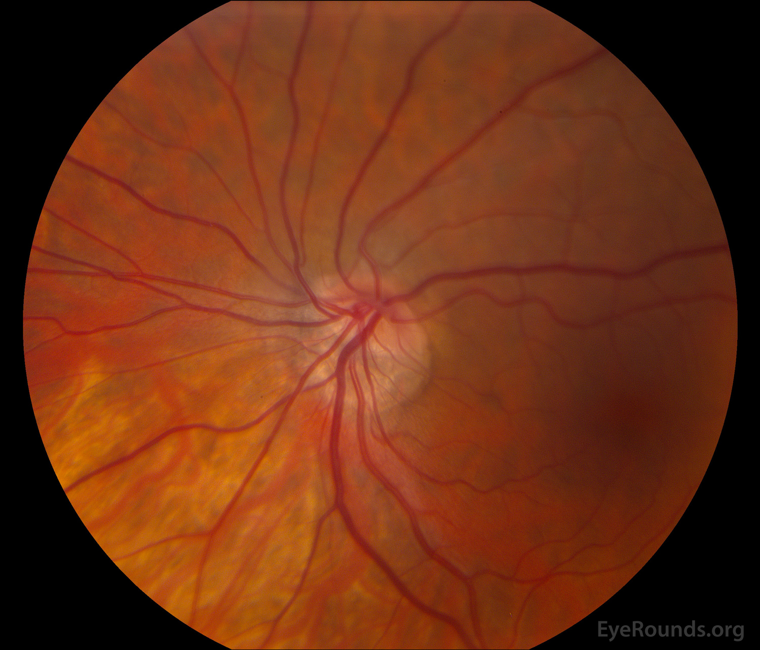
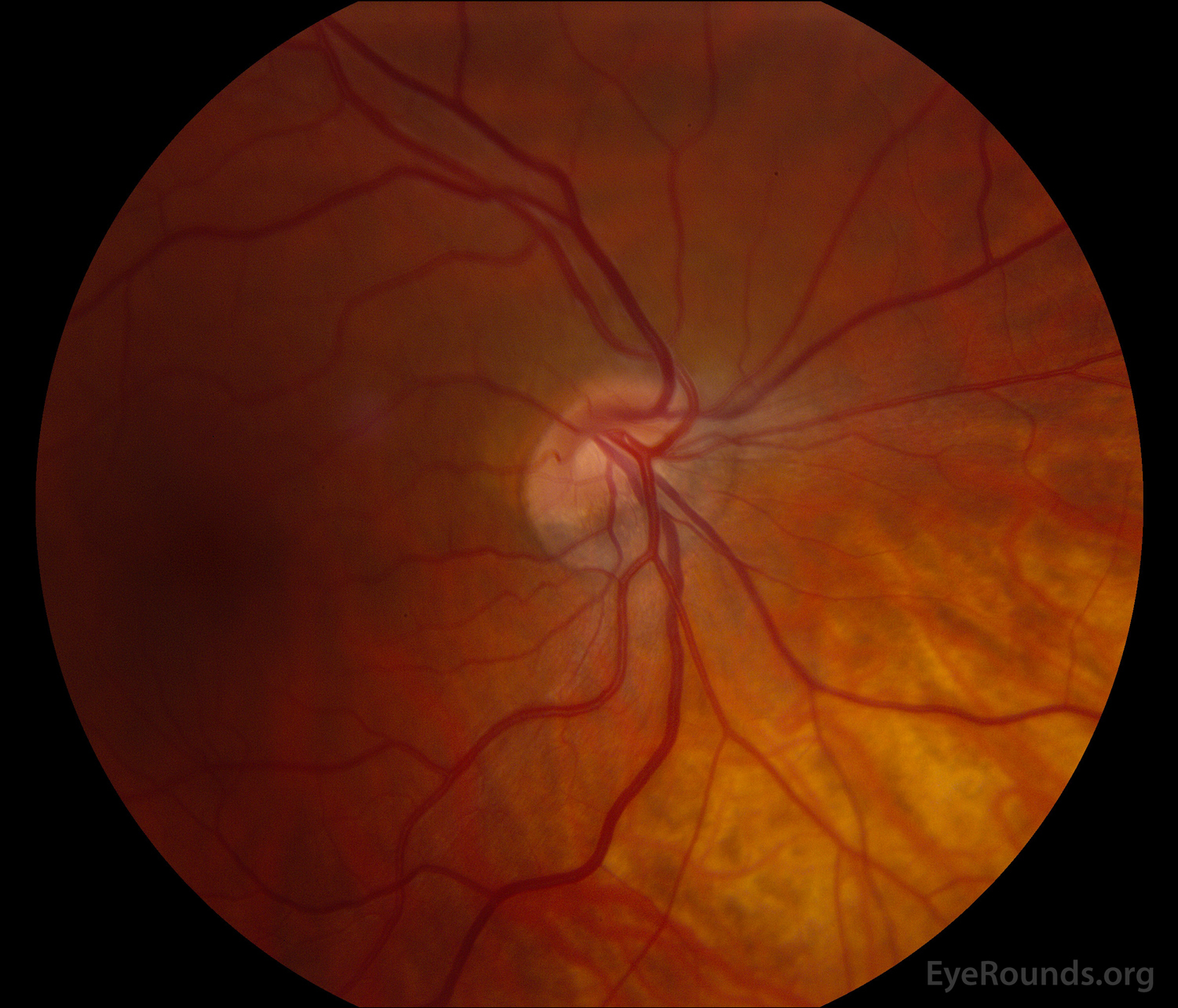
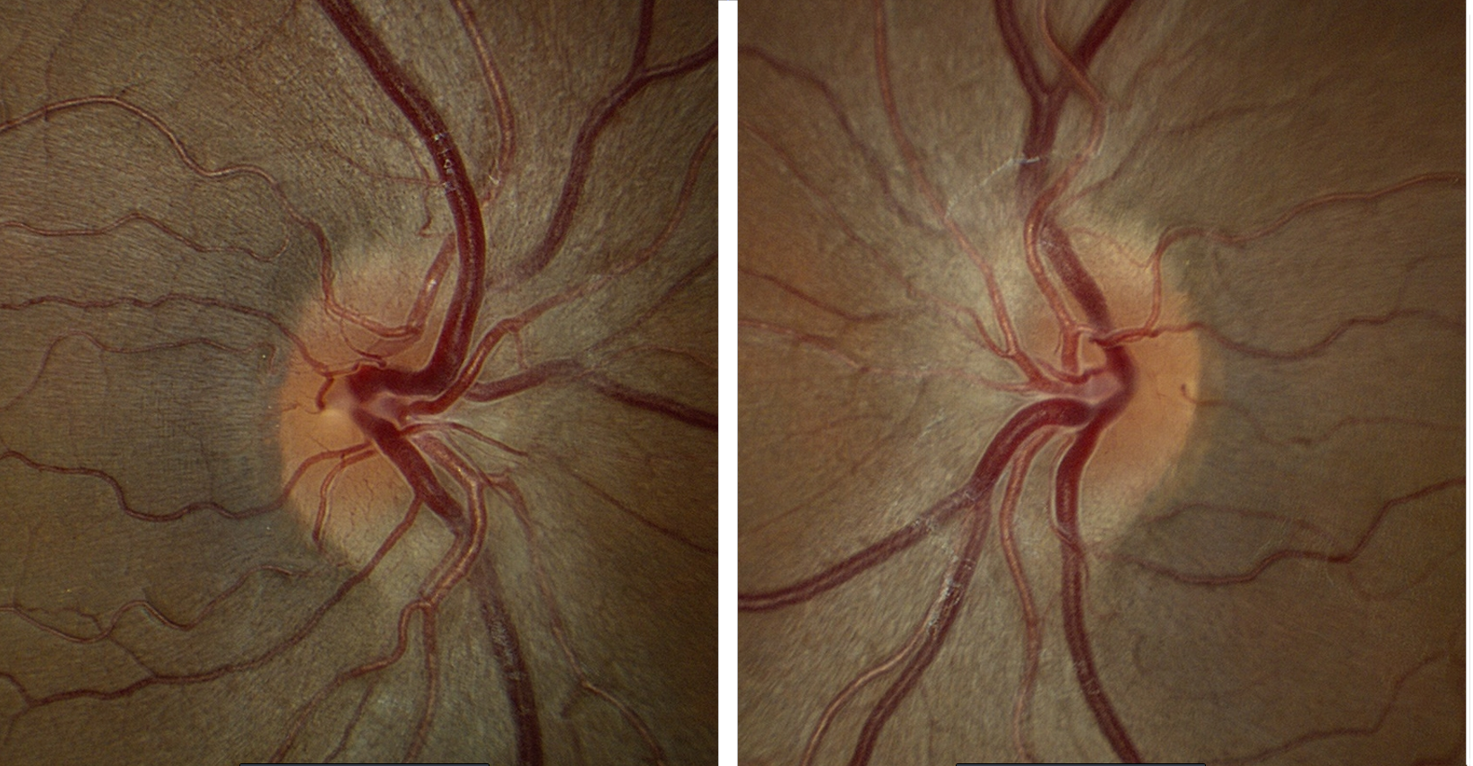
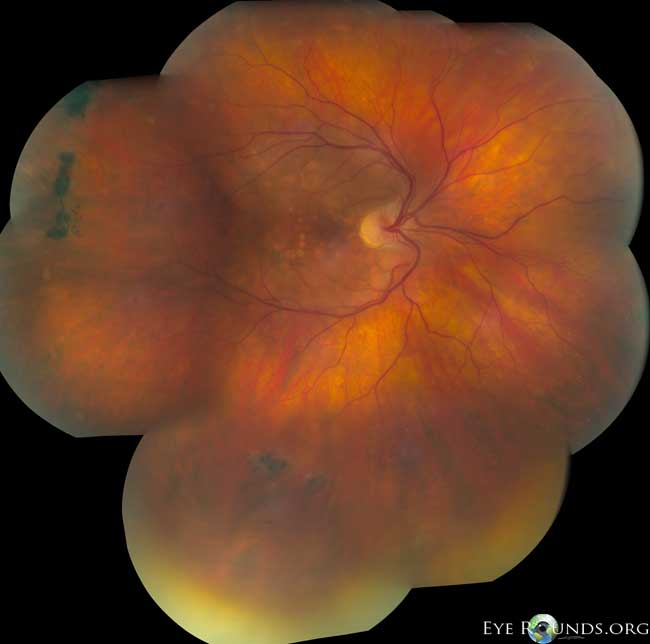
These color fundus photographs demonstrate optic nerves that are tilted to the degree of facing nasally. The retinal vessels also emerge from the nasal aspect of the disc and course nasally before turning temporally (situs inversus). The patient was asymptomatic, 20/20 in both eyes, had -8 diopters of myopia in both eyes with a normal visual field. Tilting of the optic nerve can vary in severity and is commonly associated with myopia as the optic nerve enters the sclera obliquely. Often the patient is asymptomatic, but in some cases tilted discs can be associated with superotemporal visual field defects that do not respect the midline.[1,2]
25 year-old female with congenitally tilted optic discs.
Brodsky MC. Congenital optic disc anomalies. In: Yanoff M, Duker JS, editors. Ophthalmology. 4th ed. Elsevier; 2014; Chapter 9.5; p871-874.
Taylor DS. Congenital anomalies of the optic discs. In: Lambert SR, Lyons CJ, editors. Taylor and Hoyt's Pediatric Ophthalmology and Strabismus. 5th ed. Elsevier; 2017; chapter 53; p562-580.
https://eyewiki.aao.org/Tilted_Disc_Syndrome

Ophthalmic Atlas Images by EyeRounds.org, The University of Iowa are licensed under a Creative Commons Attribution-NonCommercial-NoDerivs 3.0 Unported License.

Address
University of IowaLegal
Related Links