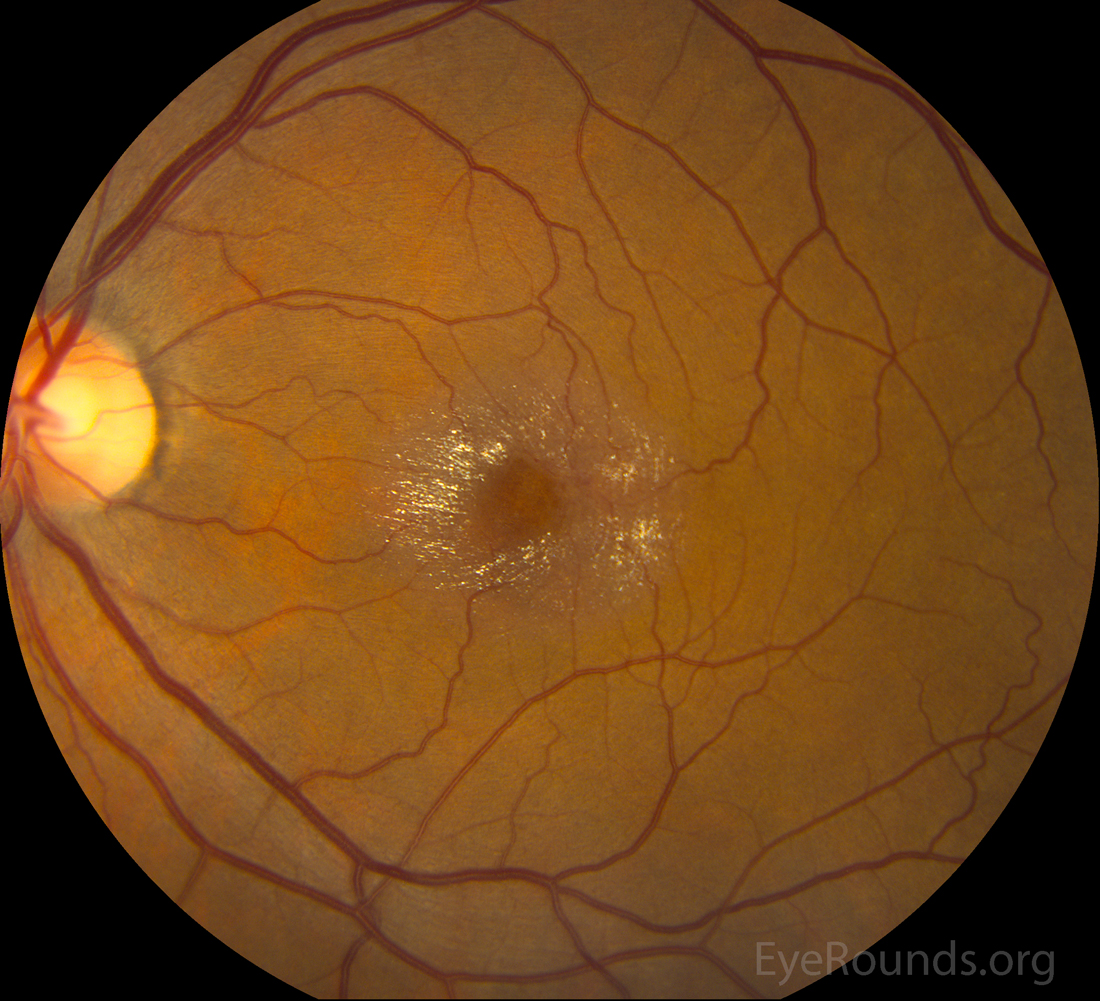
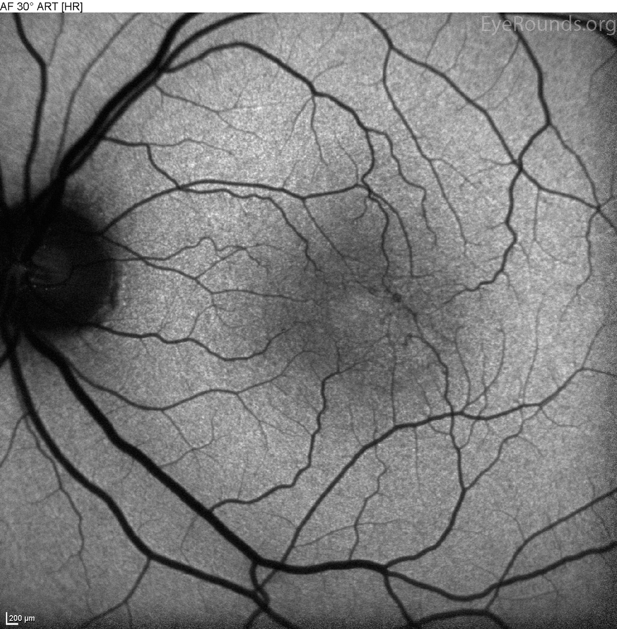
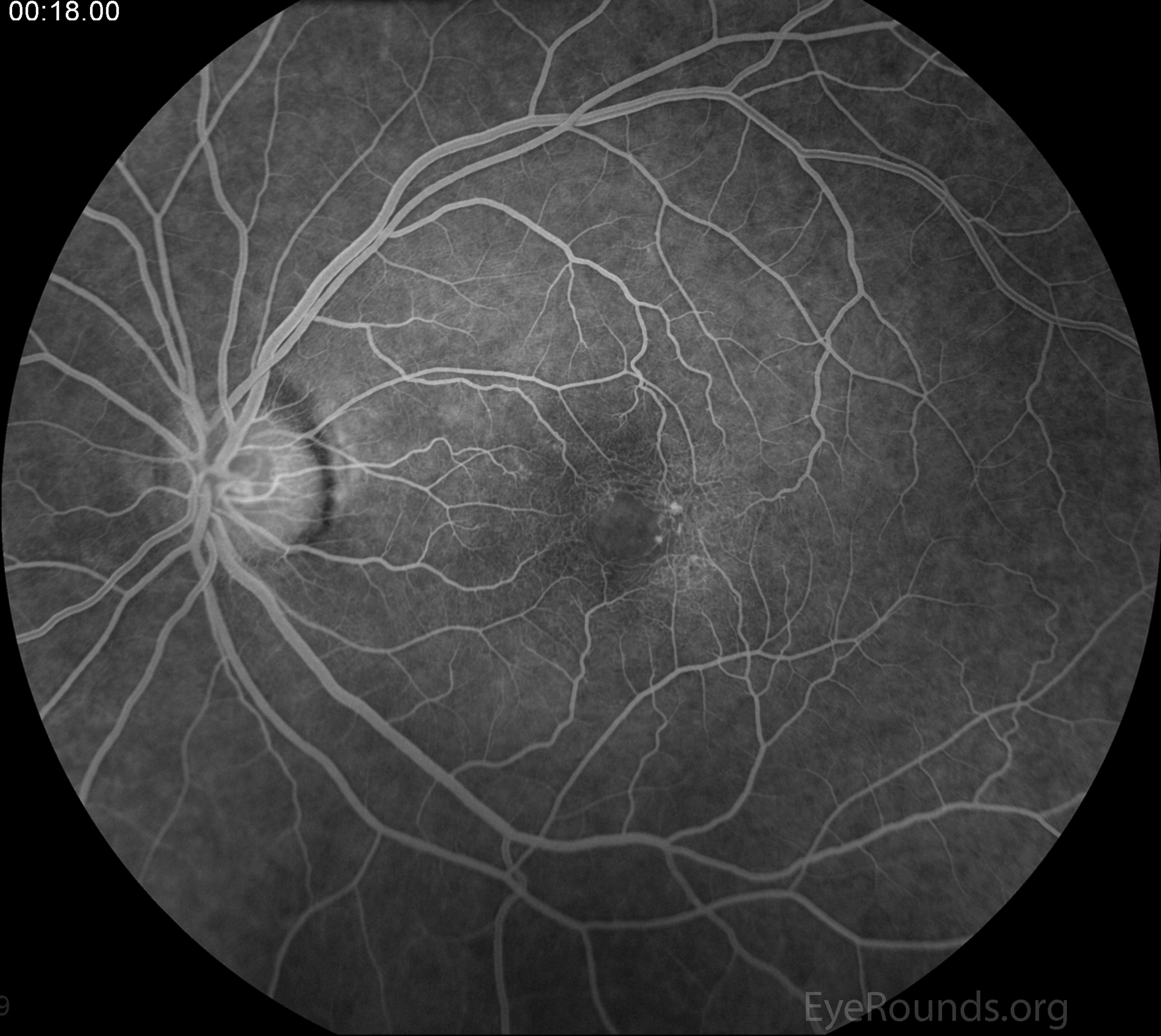
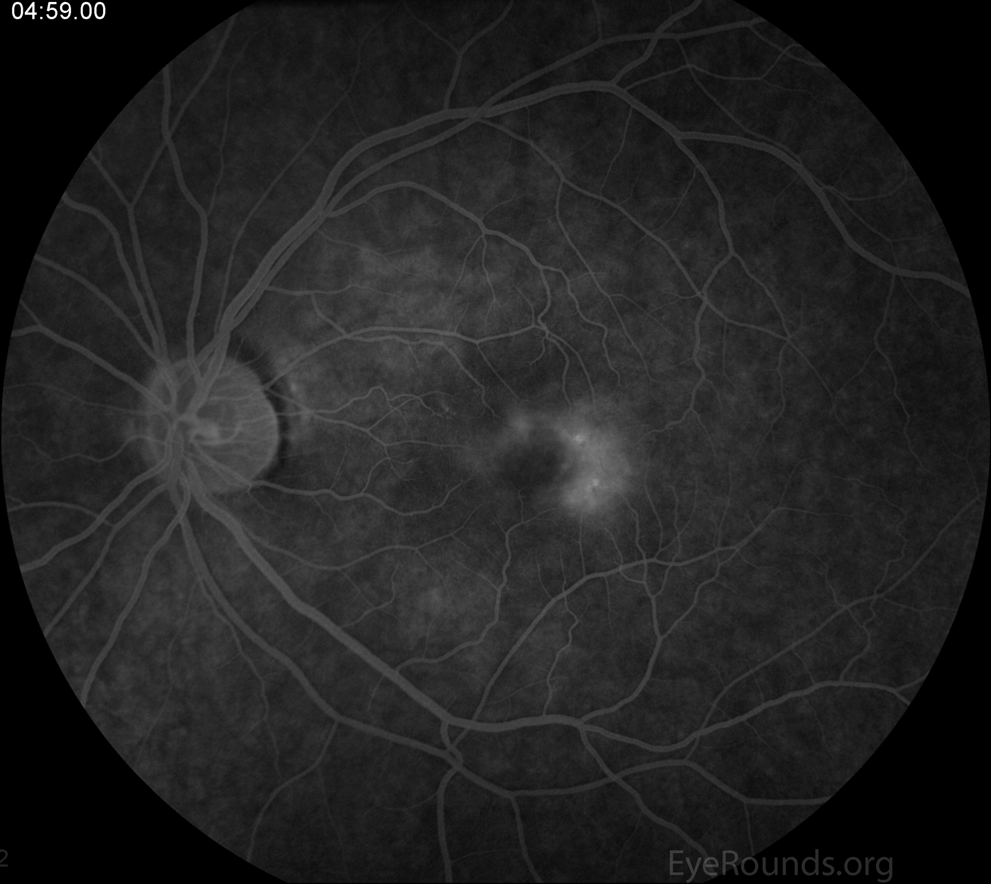
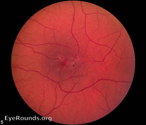
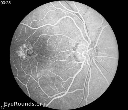
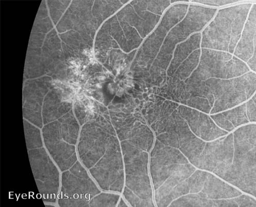
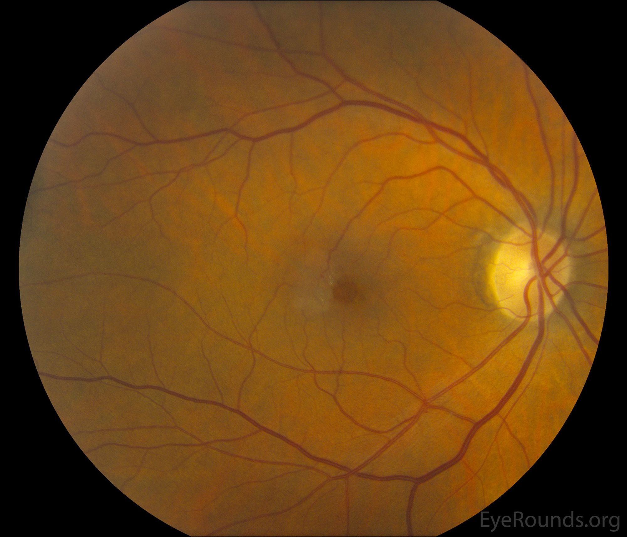
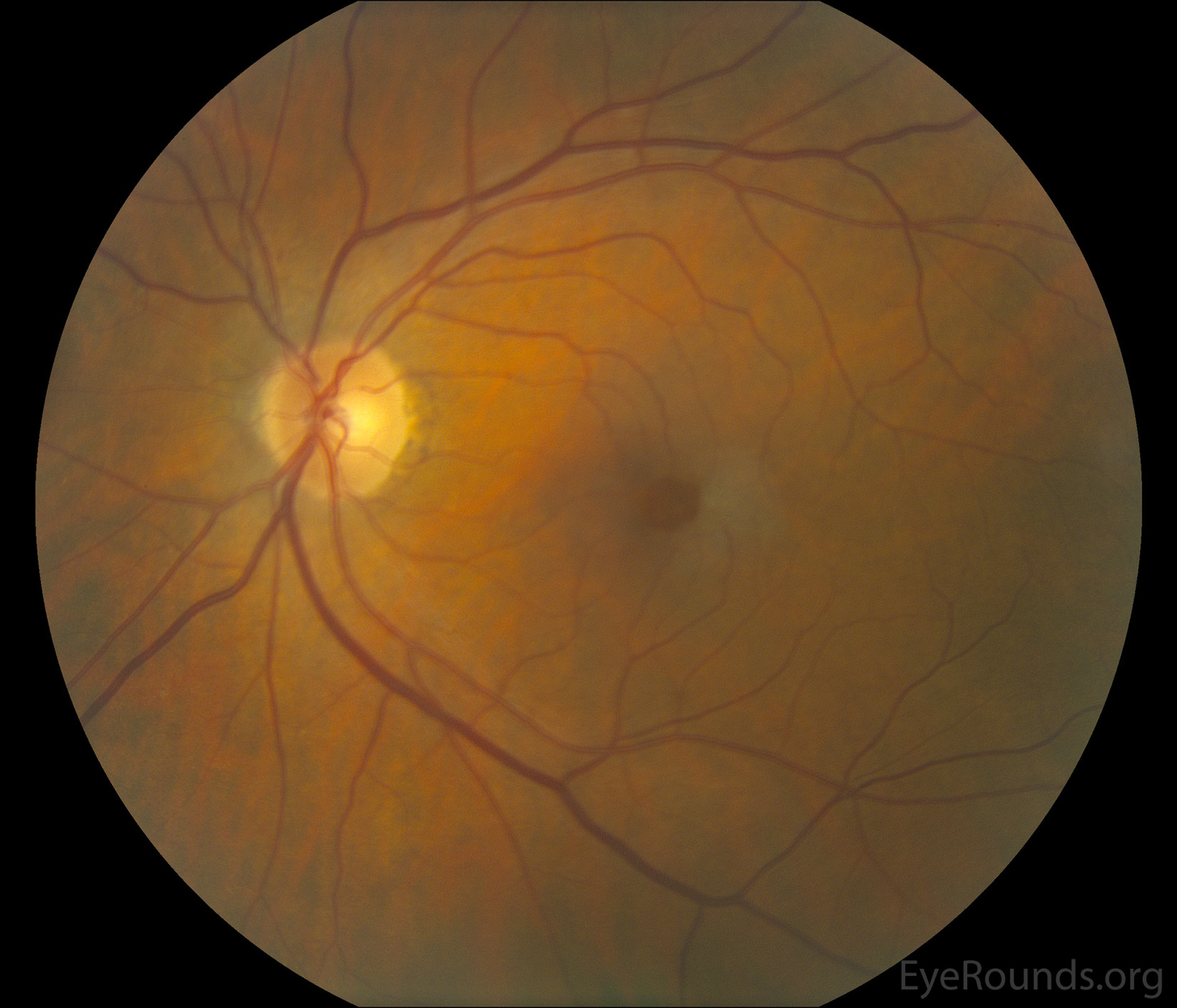
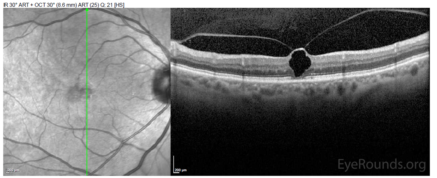
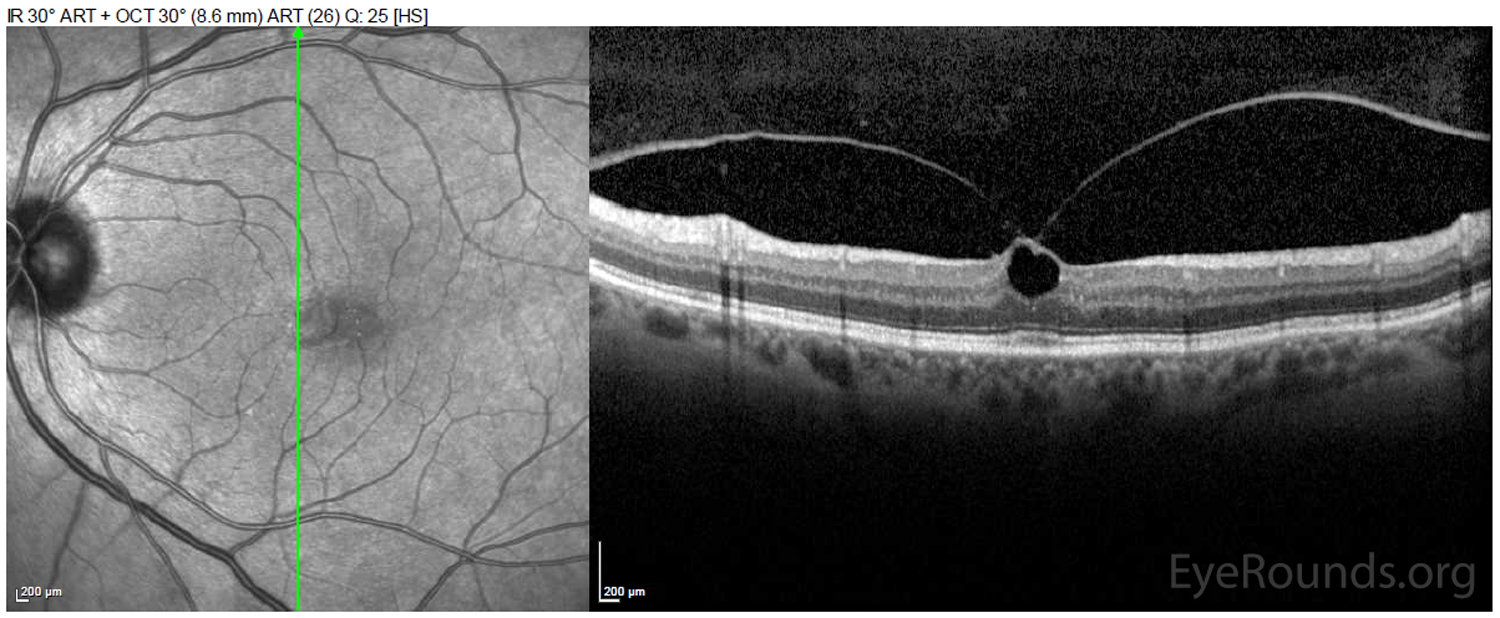
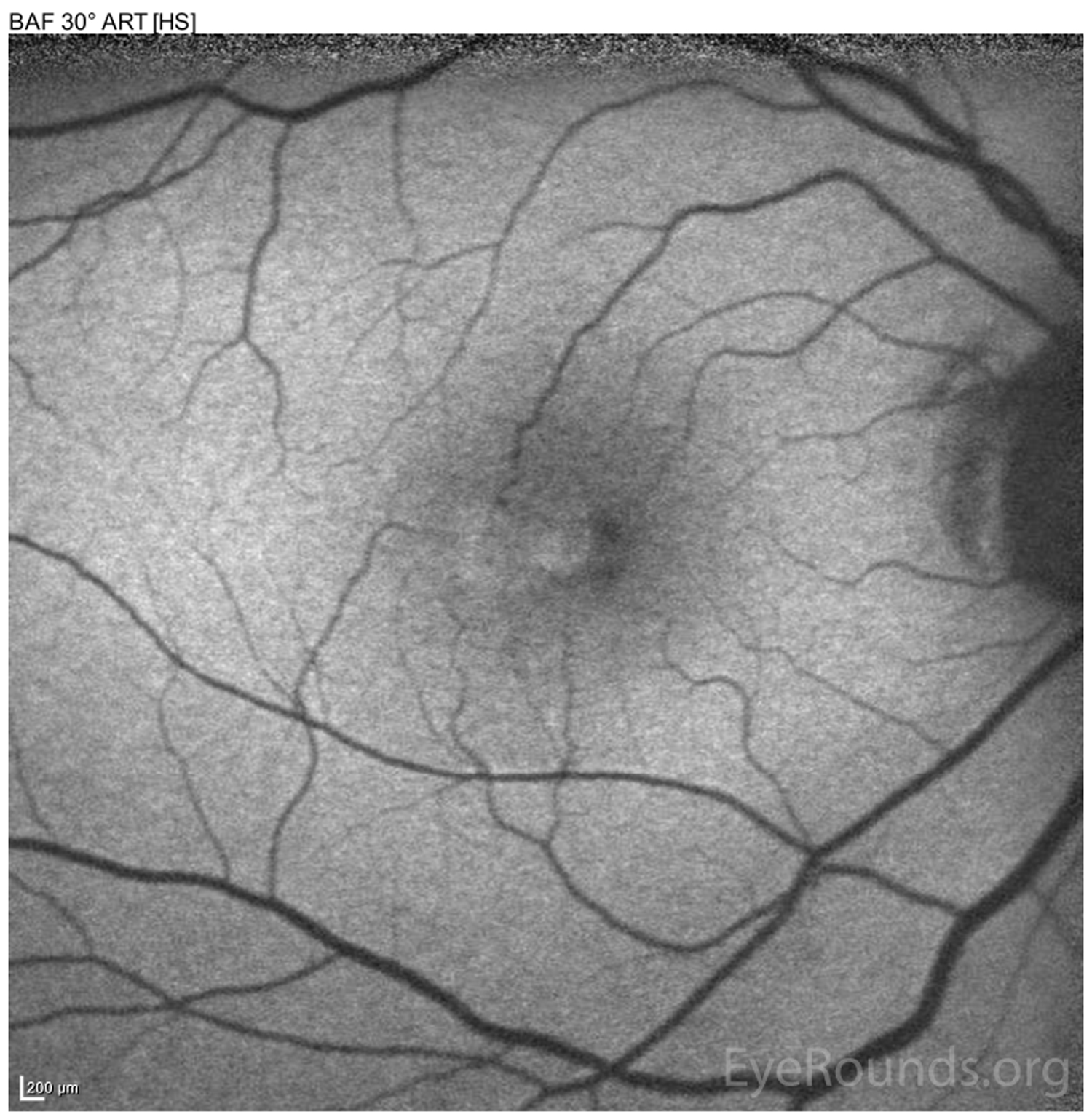
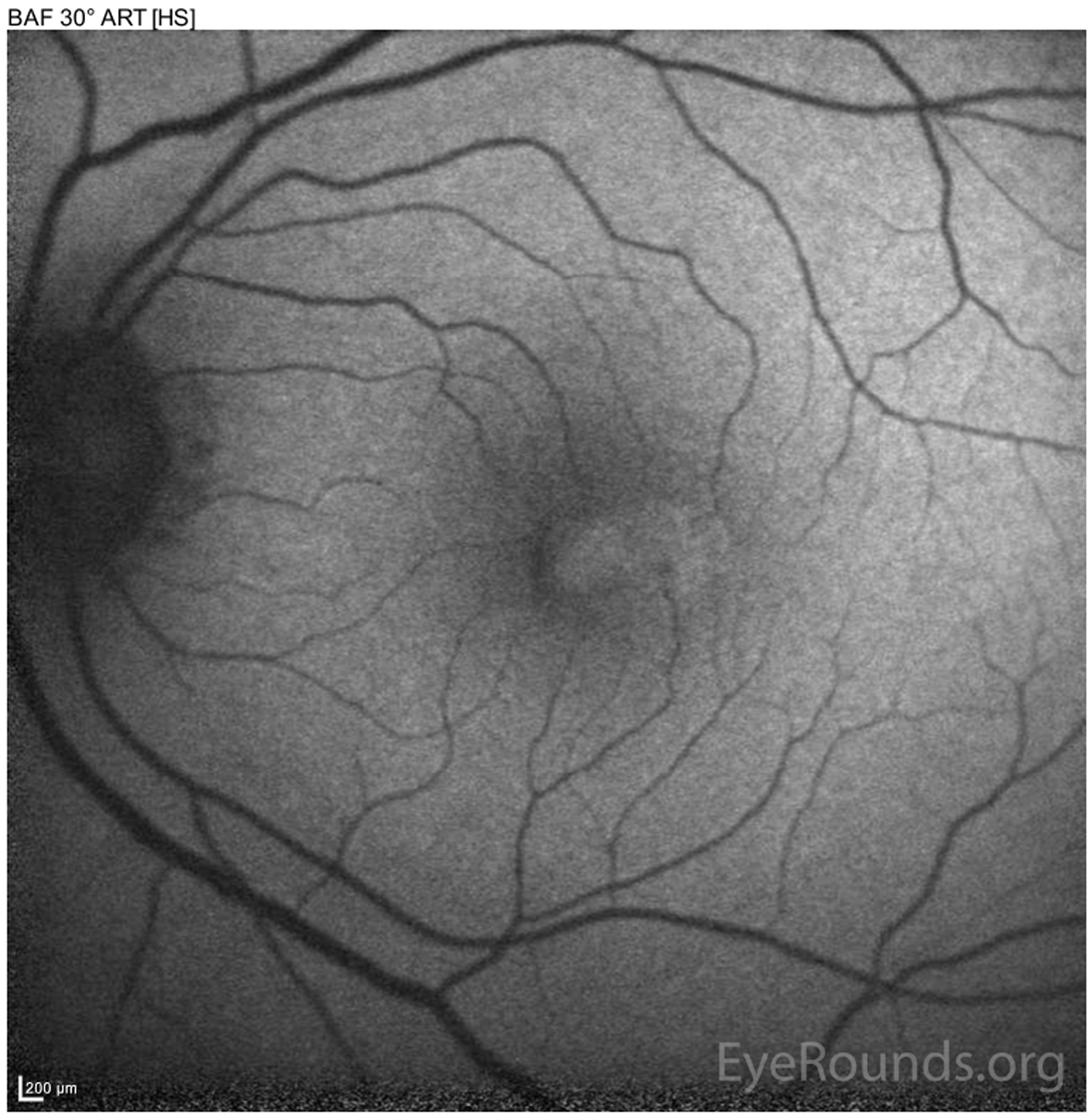
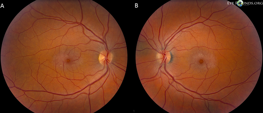
Idiopathic juxtafoveal telangiectasia type II (aka macular telangiectasia) is a rare idiopathic condition that is characterized by telangiectatic vessels in the juxtafoveolar region, most commonly temporal to the fovea, of both eyes. Additional findings include graying of the parafoveal retina, superficial crystalline deposits, subfoveal cavities, and right-angle vessels. Symptoms include blurred vision, metamorphopsia and paracentral scotomas. Fluorescein angiography will highlight the parafoveal telangiectatic vessels that show early hyperfluorescence with late leakage. OCT will show subfoveal cystoid spaces without cystoid macular edema. Fundus autofluorescence is very diagnostic in that the normal hypoautofluorescence at the fovea is lost and will show abnormally increased foveal autofluorescence. These findings are all demonstrated in the left eye of the patient shown here with very similar findings in the right eye
A 78-year-old man presented to our clinic with progressive blurring of his central vision in both eyes. Color fundus photography demonstrated a gray sheen temporal to the fovea in both eyes, which is characteristic of juxtafoveal telangiectasia (JXT). In his fovea in both eyes, there was a large, elevated round pseudo-hole due to concurrent vitreomacular traction as demonstrated by optical coherence tomography. The fundus autofluorescence images showed an absence of normal foveal hypofluorescence, replaced by an area of hyperfluorescence, a finding that is virtually pathognomonic for JXT.

Ophthalmic Atlas Images by EyeRounds.org, The University of Iowa are licensed under a Creative Commons Attribution-NonCommercial-NoDerivs 3.0 Unported License.

Address
University of IowaLegal
Related Links