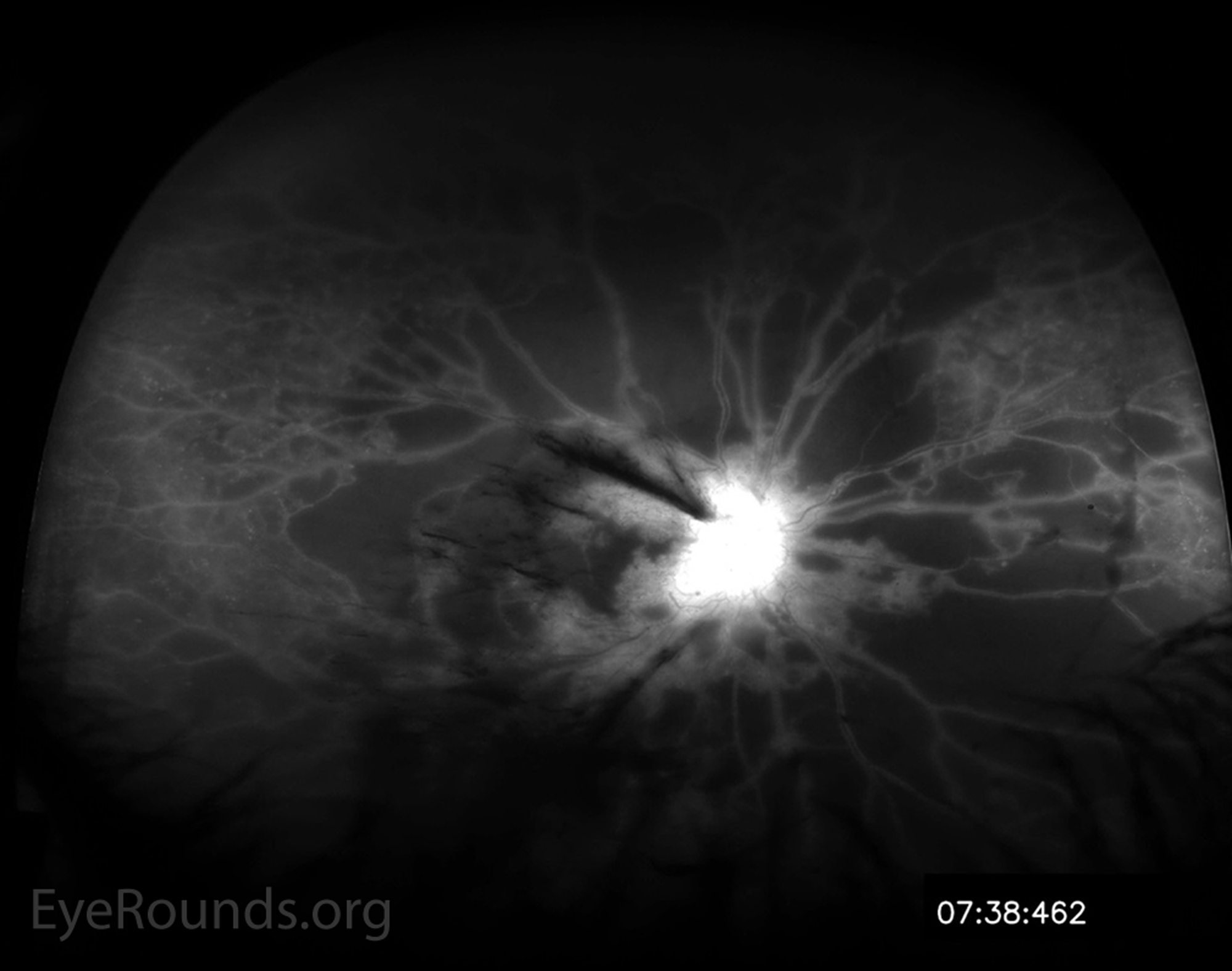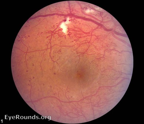
The left eye shows severe neovascularization of the disc (NVD) that extends down along the inferior arcade with tufts of neovascularization elsewhere (NVE) along both the inferior and superior arcades. The right eye had similar findings.
Posted May 11, 2017
This patient is a 32-year-old female with poorly controlled type I diabetes mellitus and severe proliferative diabetic retinopathy. She had not had panretinal photocoagulation at the time of presentation. Eventually this patient developed severe vitreous hemorrhage and traction retinal detachments in both eyes. Optos® widefield fluorescein angiography demonstrates delayed arteriovenous transit time, extensive neovascularization of the disc and huge areas of capillary nonperfusion.

February 8, 2008


Ophthalmic Atlas Images by EyeRounds.org, The University of Iowa are licensed under a Creative Commons Attribution-NonCommercial-NoDerivs 3.0 Unported License.